Health Sciences Projects
| Site: | learnonline |
| Course: | Health Sciences Projects |
| Book: | Health Sciences Projects |
| Printed by: | Guest user |
| Date: | Wednesday, 5 November 2025, 7:37 PM |
Scholarship Excellence Network
The Scholarship Excellence Network (SCENE) seeks to create a sustainable structure that utilises the knowledge and skills of successful candidates to systematically identify and support staff at scale. It is an expectation that those staff who are mentored take on the role of mentor providing sustainability and growth over time. Importantly, the adoption of a mentoring role is critical for junior staff to evidence their influence and impact for promotion purposes.
SCENE is headed by Dr Maurizio Costabile, a winner of both national and international awards. The program has begun (2019) in the Division of Health Sciences, with the support of Dean: Academic, and will also be disseminated to other Divisions for them to create a similar model.
The key outcomes of this project are:
- A Unit central point of contact.
- Pro-active identification of staff (Level A academics, casual and established) that have demonstrated excellence in teaching (through HOS, PD nominations and self-nomination).
- Facilitating the application process of eligible staff for teaching awards.
- Establishment of a pipeline of staff at varying stages in their career as potential future winners of teaching awards and grants.
For more information on SCENE, please contact Dr Maurizio Costabile.
Using 3D printed feet to teach Undergraduates Scalpeling techniques
 The project is about using 3D printed feet for training undergraduate podiatrists in scalpeling techniques, particularly for foot ulcer management.
One of the more daunting tasks for podiatry students and tutors alike is when students first begin to use scalpels on clients. This trepidation increases ten-fold when, in their later years of training, students attend a ‘high-risk’ foot clinic placement and the person who requires treatment has an existing foot ulcer that may lead to amputation if mis-managed.
Unfortunately, this is becoming more problematic with the increase in chronic disease resulting in a 30% rise in lower limb amputation rates in Australia over the last two decades. Approximately 85% of these amputations are preceded by a foot ulcer and are considered preventable with appropriate care. Scalpel debridement of the thicker callused skin that overlies foot ulcers is one of the most effective management strategies to encourage healing, yet training podiatry undergraduates in this skill is difficult due to the risks involved in allowing students to ‘practice’ on such a high-risk population. UniSA’s podiatry teaching team are tackling this with 3D printed feet and some imaginative use of icing and other moldable materials.
Drs Helen Banwell and Ryan Causby are heading up a Division of Health Sciences Dean Academic Teaching and Learning grant, along with team members Hayley Uden, Brendan Nettle and Brad Jeffery (HLS technician), to print 3D feet with various wound cavities in-situ. These cavities will be filled with various substances (officially called a ‘moulage’) to mimic non-infected and infected wounds. “Silver nitrate added to icing appears to mimic dry gangrene” says Helen. “Although we’re still working on how to make it smell realistic,” laughs Ryan.
 To support the training, ulcer debridement and management videos are being developed with the support and input of the high-risk foot clinic at the new Royal Adelaide Hospital. Once the models and videos are developed, students will be able to watch the videos and practice ulcer debridement, cleansing and dressing techniques accordingly during dedicated supervised sessions or as self-directed practice via learnonline.
The 3D models will also be used to train 2nd year podiatry students in basic scalpel handling before they begin clinical placements and 3D printed ‘baby legs’ are now being trialed in podiatry paediatric teaching sessions to allow students hands-on practice for clubfoot casting. “We estimate the models cost less than $4 to produce and, once the file is developed, can be re-printed as required” says Helen, “The possibilities are endless”. Ryan and Helen will evaluate the use of the models and the impact it has on teaching over the next 2 years.”
The project is ongoing and will expand into other areas of teaching.
If you would like to know more about the 3D feet printing project, please contact Dr Helen Banwell or Ryan Causby
Virtual Microscopy
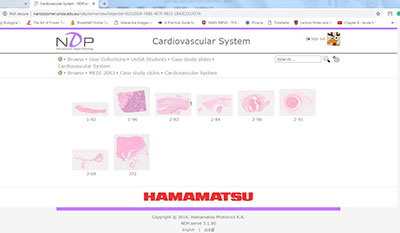 Telepathology involves the ability to capture and analyse 3D digitized micrographs of tissue sections using Virtual Microscopy (VM). Professionals and students explore and learn from whole digitized micrographs, which can be viewed at different magnifications and in different plans of field, just as when using a conventional microscope but all online. Recent advances in VM and image analysis have greatly accelerated the globalisation of pathology diagnostics and other scientific collaborations. Critically, the professional field in practice is moving in this direction.
The browser based software NDP takes the student microscope experience beyond the classroom by synthesizing microscopy technologies and digital technologies. VM circumvents traditional teaching roadblocks by removing reliance on physical space, equipment, the availability of specimens, and limited practical time. Unlike traditional sections, digital micrographs can be securely accessed online by a number of students at any time, from anywhere. Moreover, unlike standard histological slides, digital micrographs cannot be damaged during use and do not fade with time. VM enables a blended learning experience that provides students with opportunities to integrate their interdisciplinary knowledge and develop their interpretative skills. Students now have access to VM materials outside of practical sessions, promoting a deeper understanding of microscope image analysis, with the ability to collaborate with other students and staff remotely.
Exposing our students to VM has provided them with world-leading, innovative skills and a cutting-edge tool for resource sharing, networking and collaboration, and discussions have already commenced with the University of Sydney’s VM unit.
Successful integration of VM technology into the core curriculum of Pharmacy and Medical Science programs will provide a strong foundation to scale outcome beyond this initial project setting. First, the VM technology and applications will become available outside the Unit for use at both undergraduate and postgraduate levels. Second, as the university expands into online courses for students, with UniSA Online, the VM technology may be leveraged for that effort. Finally, providing students with training in VM will develop the relevant technical skills for telepathology in diagnostic and research settings that will benefit healthcare in Australia, and beyond.
Improving Teaching Quality of MBBS students
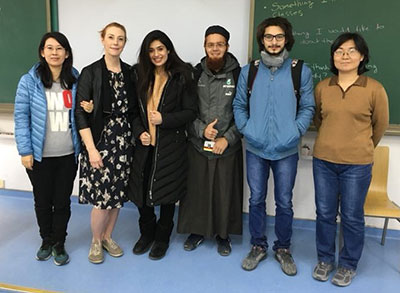 Recently, in order to improve the teaching quality of MBBS students and improve the teaching level of English, the School of Basic Medicine invited Dr. Sarah List of the University of South Australia to teach and teach MBBS students and communicate with the teachers.
Dr. Sarah List is passionate about teaching, and is good at observing students' classroom learning and focusing on interaction with students. She combines the theoretical knowledge of microbiology through specific cases and closely links the basic knowledge to clinical applications. In the teaching process, she used a variety of teaching methods such as lectures, case discussions, interactive software tests and quizzes to improve the enthusiasm of international students and achieved good learning results. Her relaxed and lively lecture style, solid professional basic theoretical knowledge, strong classroom affinity, flexible teaching methods and a smile throughout the whole process are deeply loved by international students and also a good demonstration for the college teachers.
The event was jointly promoted by the Medical Research and International Exchange Department of Qilu Medical College and the International Student Office of the Basic Medical College. The undergraduate students of the Department of Pathogenic Biology of the Basic Medical College began to select the outstanding teachers of the University of South Australia. Coordination of the content and time of the lectures, and the first demonstration project of foreign teachers in the basic medical school. This exchange program is funded by the Qilu Medical College Foreign Language Program. The School of Basic Medicine attaches great importance to the education and teaching of international students, and strives to provide exchange training opportunities for teachers in the hospital from various aspects. The development of this external teaching class project enables teachers to experience close-up experience, learn international teaching concepts and teaching methods, and enhance the international vision of teachers.
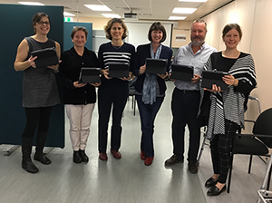
Marking Student Practical Tests using iPad marking system
Our project is about improving the quality and the ease of marking student practical tests by using an electronic marking system on an iPad instead of pen and paper.
The process we used for marking practical skills tests in our physiotherapy program was not working well for students or staff. The traditional pen & paper way of marking was time consuming to do during the test, errors occurred when transcribing the marks from paper to the University online system, it was time consuming for staff to add up, collate and moderate student marks across large class sizes, and time consuming to provide individualised written feedback which students had to then come in to campus to collect a couple of weeks later, with many students not collecting and using their feedback. As University classes are increasing in size and more students have the opportunity to attend University here at UniSA, the time spent on practical assessments is increasing yet the same issues exist. So a team of us here in our Unit sought an improvement to the process. We got in touch with a physiotherapy academic at Curtin University, Dr Leo Ng (ing), who had presented a new iPad assessment tool at an industry conference. And so a collaboration began.
The project has been very successful. It has been trialed in 7 Physiotherapy courses and one Podiatry course in 2018. The trial involved over 450 students and 19 staff assessors. The feedback we gained from staff and students was overwhelmingly positive. Students like their individualised, detailed feedback provided within days of their test via email, including images of their performance. Staff like the ease of using the iPads, the automatically produced student feedback, and the large time savings to complete and moderate practical assessments.
 Key to the success was the willingness of the iPad marking system developers to share the technology, the flexibility of the system to be adapted to meet our specific needs here at UniSA, the expertise and enthusiasm of the project team members to conduct the trial, the financial support of the university, and the willingness of Physiotherapy and Podiatry staff and students to trial the new technology.
With the support of the Division, both Physiotherapy and Podiatry programs will be continuing to use the iPad marking system in 2019. And we will be able to share the technology with other interested programs in the Division of Health Sciences including Sonography and Exercise and Sports Science to improve the practical assessment experience for more students and staff..
If you would like to know more about the iPad marking project, please contact Dr Caroline Fryer.