Micrometry
Micrometry refers to the measurement of the size of particles examined in the microscope. In order to be able to estimate the size of particles seen in the microscope, a point of reference is needed as microscopes are not necessarily uniform in their optical properties. An ocular graticule is used to actually measure the particles or cells, but this needs to be calibrated for each microscope and each objective. Graticules are placed into an ocular lens and simply consist of some regularly spaced ruled lines. They are calibrated by observing a stage micrometer slide or haemocytometer through the graticule.
CALIBRATION
- Focus the ruled lines of the graticule and the ruled lines on the micrometer or haemocytometer simultaneously.
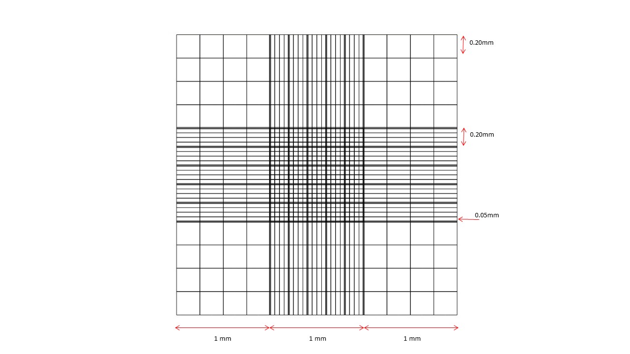
- Move the stage so that the graticule corresponds to a known graduation in the micrometer or haemocytometer, e.g 10 graticule lines corresponding to the distance between the closest lines in the haemocytometer. Sizes for the rulings in the haemocytometer are shown below. In this case, 1 graticule unit would therefore equal 0.05mm or 50 microns.
- Repeat the calibration for each objective (except the 100x).
USE OF THE OCULAR GRATICULE
- Observe the specimen through the ocular graticule, positioning the object such that it lies between the ruled lines.
- Determine how many graduations correspond to the object.
- By reference to the calibrations, determine the size of the object. If the object is not round or square, measure both the long and short axes.
- For very small objects such as bacteria, try to measure a number of cells attached to each other e.g. if 5 cells fit between each graduation, divide the size of the graduation by 5 to give the size of the object.
- For accuracy take a number of measurements (5-10) and determine the mean.
NEUBAUER COUNTING CHAMBER
Last modified: Monday, 5 February 2018, 10:27 AM