Enumeration
On this page:
INTRODUCTION
In order to be able to construct a growth curve, and for other applications such as antibiotic sensitivity testing, a means of calculating the numbers of bacteria in a culture is essential. Cultures of bacteria always contain a mixture of living and dead cells, in the log phase of growth the majority of cells are living (viable) but after the stationary phase, many are dead. Total numbers can be estimated from the turbidity of a culture and during log phase there is a direct correlation between viable count and turbidity. This correlation breaks down as numbers of dead cells increases.
MEASUREMENTS OF TURBIDITY
NEPHELOMETRY
The light from a standard source is directed onto the suspension of bacteria in a cuvette. Particles (bacteria) in the suspension will scatter the light and the amount of scattering is proportional to the numbers of particles. Light scattered at right angles to the original light path is detected by a photoelectric cell and registered as a deflection on a galvanometer. The scale on the galvanometer is arbitrary and must be calibrated against known concentrations of bacteria. The calibration must be performed for different types of organisms as light scattering is different, for example, for cocci versus rods.
Simple nephelometry is useful for estimating the turbidity of a culture prior to antibiotic sensitivity testing, where the turbidity can be adjusted to a standard preset value.
OPACITY TUBES
This method consists of matching by eye the opacity of a bacterial culture with a set of prepared standards containing BaSO4 in a finely divided suspension at different concentrations. The concentration of the BaSO4 standards is calibrated against known concentrations of bacteria in a nephelometer such that each tube corresponds closely to a set concentration of bacteria. An example of these tubes are McFarland standards, prepared as follows:
| Tube number | Barium chloride 1% w/v (ml) | Sulphuric acid 1% w/v (ml) | Density of bacteria (x106/ml) |
| 1 | 0.1 | 9.9 | 300 |
| 2 | 0.2 | 9.8 | 600 |
| 3 | 0.3 | 9.7 | 900 |
| 4 | 0.4 | 9.6 | 1200 |
| 5 | 0.5 | 9.5 | 1500 |
| 6 | 0.6 | 9.4 | 1800 |
| 7 | 0.7 | 9.3 | 2100 |
| 8 | 0.8 | 9.2 | 2400 |
| 9 | 0.9 | 9.1 | 2700 |
| 10 | 1.0 | 9.0 | 3000 |
The tubes are sealed to prevent evaporation.
USE OF McFARLAND STANDARDS
- Place approximately 1 ml of the culture to be measured into a glass tube of the same diameter as the standard tubes.
- Resuspend the standards thoroughly.
- Hold the tube between successive pairs of the standards e.g. pick tube number 1, then your sample and then tube number 2.
- Compare the opacity by holding the tubes over the card ruled with thick black lines (zebra paper) supplied. Alternatively, hold the tubes over some bold printing in your practical books.
- Determine the tube number which matches the sample most closely.
- Read the estimated concentration of bacteria from the table on the end of the rack of standards. Remember that the values are multiplied by 106. For example, if the culture corresponds to tube 3, the concentration is 900x106 bacteria/ml which is equivalent to 9x10⁸ bacteria/ml. (See the chapter on laboratory calculations for ways of dealing with very large numbers.) This concentration (9x10⁸ bacteria/ml) is commonly achieved in an overnight culture of E. coli or S.aureus and hence these large numbers must frequently be dealt with in Microbiology.
Use of MacFarland opacity standards video
SPECTROPHOTOMETER
A reasonably accurate measure of the number of bacteria present in a suspension can be obtained by measuring the optical density. This gives a similar count to the nephelometer and must also be calibrated for each organism. The optical density is measured at 550 μm after zeroing the machine against the medium in which the organisms are suspended.
MANUAL COUNTING METHODS
These counting methods are usually used to count cells such as red blood cells and various white blood cells and are not as accurate when used to enumerate bacteria. However, an estimate of bacterial numbers can be made if a suitable suspending fluid is used to produce adequate contrast or a phase contrast microscope is used. Bacterial cultures generally have too many cells to be able to be counted directly, so a dilution into a suitable contrast enhancing solution is employed. Inclusion of formalin in the solution fixes the bacteria and ensures motile organisms are not moving, which can make the count inaccurate.
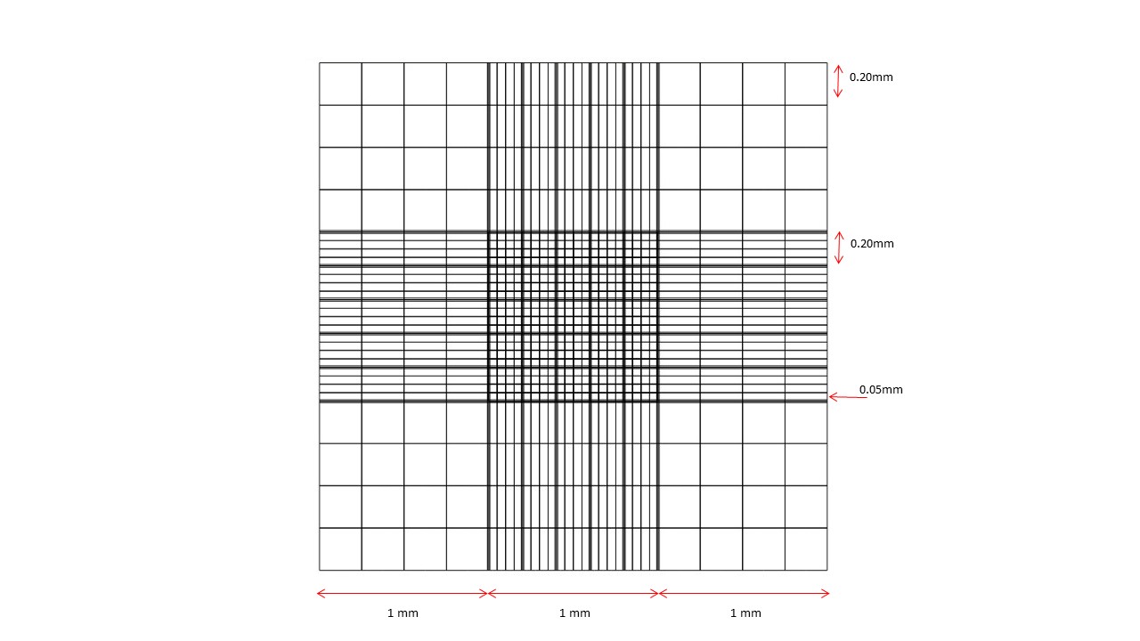
NEUBAUER HAEMOCYTOMETER
The grid of the Neubauer haemocytometer chamber is pictured below, with the sizes of the squares. The depth of the chamber when filled is 0.1mm. Hence the volume of the smallest square in the grid is 0.05x0.05x0.1 mm3 which is equivalent to 0.25x10⁻⁶ ml.
PROCEDURE FOR BACTERIAL COUNTS
Diluting fluid:
- Freshly filtered Loeffler methylene blue 1 vol
- Formalin 1 vol
- Saline 18 vols
Procedure:
- Dilute the culture 1 in 20 with the diluting fluid.
- Fill both sides of the chamber with a Pasteur pipette. Do not overfill as the volume will be incorrect.
- Using the 40x objective, count the numbers of bacteria in 20-50 of the smallest squares in the centre of the grid.
- Calculate the average number of bacteria per smallest square.
- Calculate the concentration of bacteria in the ORIGINAL sample by:
Number per ml = ave. number per small square
dilution factor x vol. small square
= ave. number per small square
1/20 x 0.25x 10-6
= ave. number per small square x 8 x 10⁷
KOVA GLASSTIC SLIDE
The Kova slide is used to determine the number of eukaryotic cells in a suspension; for example, the numbers of red or white blood cells in a urine specimen or the numbers of cells in a tissue culture. It is not accurate for bacterial counts as the squares are too large.
PROCEDURE
This is used to count the number of cells of patient origin (polymorphonuclear cells, red blood cells, epithelial cells etc) or tissue culture cells and not numbers of bacteria.
- Mix the specimen well.
- Draw up some suspension in a pasteur pipette, hold the pipette tip in the notch on the slide and gently expel a drop of liquid into the chamber. Try not to overfill.
- Count the number of each type of cell in 10 small squares if there are many cells, or in the whole grid if there are only a few cells.
- Calculate the number of each cell type per ml by the following formula : number = Average number per smallest square x 90 x 103/ml.
VIABLE COUNTS
The principle of the viable count relies on the development of a colony on a solid medium or growth in a liquid medium from a single viable cell. The assumption is made that a single colony is the result of a single cell but in practice, 2-3 cells in close proximity may give rise to a single colony. This problem is difficult to eliminate but can be minimized by using a thoroughly mixed suspension and not crowding the plate with too many cells/colonies. As such the suspension of bacteria will need to be diluted, usually in a series of 10 fold dilutions, but occasionally in a series of doubling dilutions (i.e. 2-fold dilutions).
- 10-fold serial dilutions give rise to the dilutions 100(undiluted),10-1(1/10),10-2(1/100),10-3 (1/1000) etc.
- 2-fold serial dilutions give rise to the dilutions 20,2-1(1/2),2-2(1/4),2-3 (1/8) etc.
SIMPLE VIABLE COUNT PROCEDURE
Each dilution may be plated to produce duplicates or triplicates where appropriate.
- Add 900μl of sterile saline to a number of sterile eppendorf tubes (see notes).
- With an autopipette, add 100 μl of the bacterial culture to a nutrient agar plate and spread with a spreader.
- Using the same pipette tip, add 100 μl of well mixed bacterial suspension to the first tube of saline (10-1). Mix well.
- Using a fresh tip, remove 100 μl from the 10-1 tube and add to the next agar plate and add another 100 μl to the next tube (10-2). Mix well.
- Using a fresh tip, remove 100 μl from the 10-2 tube and add to the next agar plate and add another 100 μl to the next tube (10-3). Mix well.
- Repeat for all dilutions required.
- Incubate the agar plates appropriately.
- Count the colonies on the plates containing between 30 and 300 colonies.
- Applying the appropriate dilution factor, calculate the number of viable bacteria in the original culture:
Example:
57 colonies counted from 0.1 ml placed on the 10-5 plate
Concentration = 57 x 10 x 105
= 5.7 x 10⁷ bacteria/ml
NOTES
- The number of dilutions required will depend on the initial concentration of bacteria in the suspension, which can be estimated from turbidity measurements (e.g. McFarland tubes). Dilutions must then be prepared to cover concentrations both above and below the estimated concentration.
Example
McFarland estimate = 6x10⁸ bacteria/ml
This concentration would give 600 colonies on the 10-5 plate (too many) and 60 on the 10-6 plate, so that dilution to at least 10-7 is required to allow for errors in the estimate. Some of the lower dilutions (10-1-10-4) may be omitted from plating. - For most purposes, nutrient agar is suitable but other non-selective media can be used. Selective media are not suitable as they may be partially inhibitory to the organism and an underestimate will result.
- Dilutions can be prepared with different starting volumes as required. Volumes less than 100μl can be conveniently prepared in microtitre trays.
MOST PROBABLE NUMBER (MPN) COUNTS
This procedure is used extensively in the food industry and in testing water quality where numbers of bacteria in the material to be tested are expected to be low. In the food and water industries, the organisms of interest are predominantly coliforms (gram negative rods related to E. coli) and the medium into which the samples are introduced/diluted is often a medium which acts to identify the bacteria as well as allowing their growth.
MPN counts are essentially a statistical method. In essence, the material is diluted to a point where the inocula may sometimes but not always contain a viable organism. The numbers of inocula at a given dilution that produce growth give an estimate of the original concentration (by reference to standard tables).
The technique can be modified to sample fluids (e.g. milk or water) or solid foodstuffs. A series of 3 consecutive decimal dilutions is chosen and 3, 5 or 10 replicates are made for each dilution. For solids, a series of 3, 10-fold reductions in weight are added to the broth e.g. 10g, 1g and 0.1g instead of a dilution of liquid. In the lower dilutions (1/10) and larger masses, all tubes are expected to grow; while in the higher dilutions or smaller masses, no tubes may grow. From those dilutions/masses in between, some replicates may grow and others not for a given dilution/mass. By reference to the standard tables, the most probable number of organisms in the original sample can be determined, depending on how many growths/no growths are seen. The more replicates, the more accurate the estimate; however, it is not always practical or cost effective to inoculate 10 tubes for each dilution.
EXAMPLE
10, 1 and 0.1g of minced beef are added to 3 tubes each of lauryl sulphate tryptose broth (for the growth of coliforms). After incubation, 3 tubes from the 10g set showed growth of coliforms, 2 from the 1g set and 0 from the 0.1g set. Referring to 3 tube tables (see the first reference below), the MPN of coliforms in the sample is 0.93/g.
In practice, these tests are more complex because of the necessity to confirm the presence of coliforms with further tests.
REFERENCES
Reasonably straightforward explanation of the technique can be found here:
https://microbeonline.com/probable-number-mpn-test-principle-procedure-results/
Other references:
American Public Health Association. Standard Method 9221C. Estimation of Bacterial Density. (1992) in Greedberg AE, Clesceri LS, and Eaton AD. Standard Methods for the Examination of Water and Wastewater. 18th Ed.
Oblinger JL and Koburger JA (1975) J. Milk Food Tech. 38:540-545
De Man JC (1983) Eur. J. Appl. Biotech. 17:301-305 (standard tables)
MPN Table Calculator:
http://www.i2workout.com/mcuriale/mpn/