18961 NEUROBLASTOMA ADJACENT TO THE HEAD OF THE PANCREAS
The patient was a man aged 62 who presented with vomiting and progressive jaundice. A presumptive diagnosis of carcinoma of the pancreas was made and appeared to be confirmed at laparotomy. Gastro-enterostomy and cholecystojejunostomy were performed. After the operation there was respiratory difficulty, necessitating incubation and treatment on the respirator, and he was anuric. On the 5th day he developed high fever and died. At postmortem secondary deposits were found in the mediastinal and para-aortic lymph nodes, the liver, the left adrenal, the mesentery, omentum and perinephric fat.
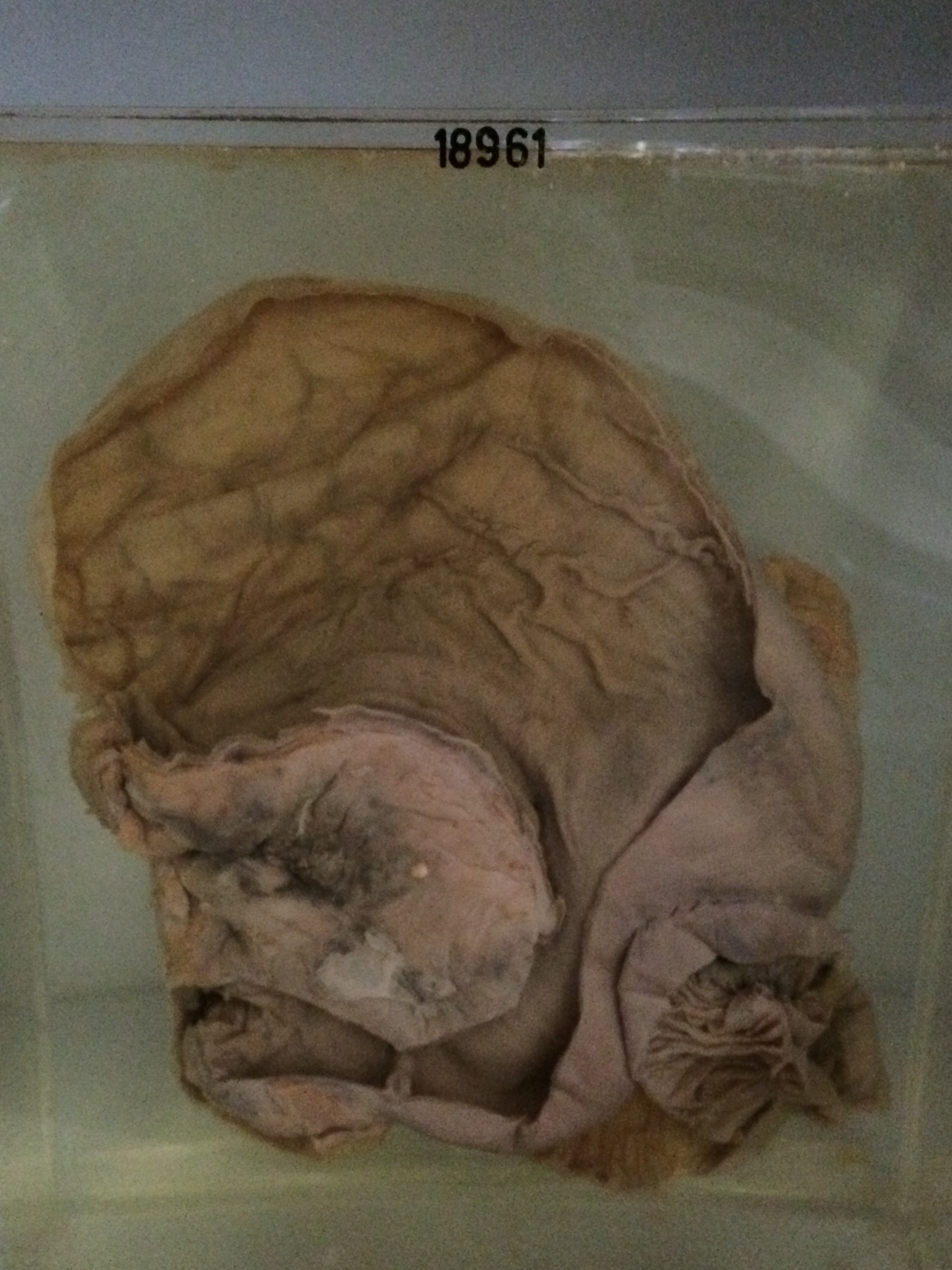
The specimen consists of the stomach, duodenum and pancreas. The stomach is dilated and shows evidence of chronic hypertrophic gastritis in the pyloric half. The termination of the first part of the duodenum and the 2nd part of the duodenum are distorted by a tumour in the region of the head and body of the pancreas which is ulcerating into the left side of the 2nd part of the duodenum. The gastroenterostomy is intact. A large cyst of the lesser sac is visible on the reverse of the specimen. It lies below the tail of the pancreas and measures about 8 x 7 x 6 cms. This cyst contains old blood and fibrinous exudate. There is a second similar smaller cyst above it. The tumour has infiltrated directly into the lesser omentum and around the margins of the foramen of Winslow. The common bile duct was obstructed by tumour infiltrating and compressing its wall. Histology shows a mass of small carrot-shaped cells with regular ovoid nuclei and scanty cytoplasm. Rosettes are visible in some places. A small nodule of tumour is present in the left adrenal, which is otherwise normal. There is no tumour in the wall of the cyst in the lesser sac. The tumour presumably arose from a sympathetic ganglion of the paravertebral chain. It is a rare tumour, especially in the elderly.