15957 CHRONIC PULMONARY TUBERCULOSIS AND PRIMARY CARCINOMA
This patient was a man aged 55 with a 12 year history of progressive fibro-caseous pulmonary tuberculosis. For the last 5 years no bacteria had been demonstrable in the sputum. For 6 months before admission he had complained of pain in the 5th right intercostal space. For the last month there was a rash in the right 9th and 10th thoracic dermatomes, considered to be herpes zoster. Recent X-rays showed rapid extension of an opacity in the right midzone which was thought to be a carcinoma. He was admitted to hospital for investigation but died after one week. At postmortem there was fibro-caseous tuberculosis of both upper lobes and a primary carcinoma in the right lower lobe. No secondary deposits were apparent anywhere in the body, including the mediastinum.
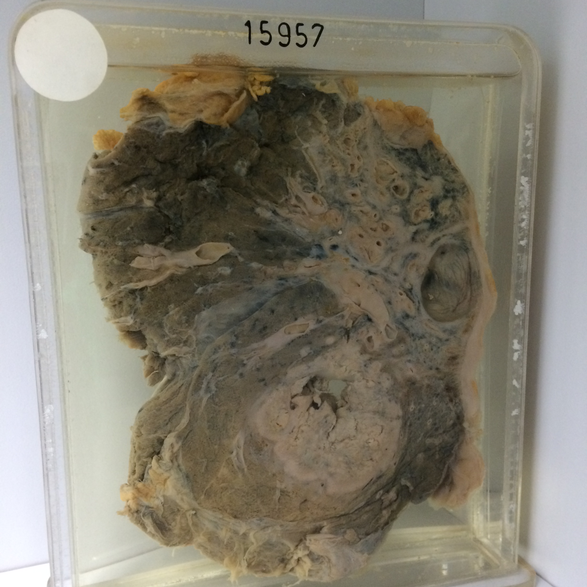
The specimen is the right lung sectioned to show dense fibro-caseous tuberculosis with bronchiectasis in the upper lobe, in an area about 6 cms in diameter where dilated bronchi lie in a dense mass of pigmented fibrous tissue. A thin-walled cavity 2.5 cms in diameter is present just beneath the pleura in the apical segment of the lower lobe. There is a primary carcinoma 5 cms in diameter in the substance of the lower lobe. It shows a good deal of central necrosis and cavitation and there is some ill-defined pneumonic consolidation in the surrounding lung. Histology shows that the apical cavity is partly epithelialised without evidence of active tuberculosis in its walls. The adjacent areas are very densely fibrous and contain dilated bronchi with normal epithelium and small cystic spaces filled with foamy macrophages. The tumour is an actively growing epidermoid carcinoma.