18378 LOBAR PNEUMONIA
The patient was a man aged 85 who had congestive cardiac failure. He was admitted to hospital in acute respiratory distress, with pain in the chest made worse by breathing. There was auricular fibrillation and signs of acute pulmonary oedema. The JVP was raised. He died 8 hours after admission. Postmortem showed massive pneumonia in the right lung and a smaller similar area of pneumonia in the left lung. The right pleural cavity contained about 4 ozs of turbid fluid exudate.
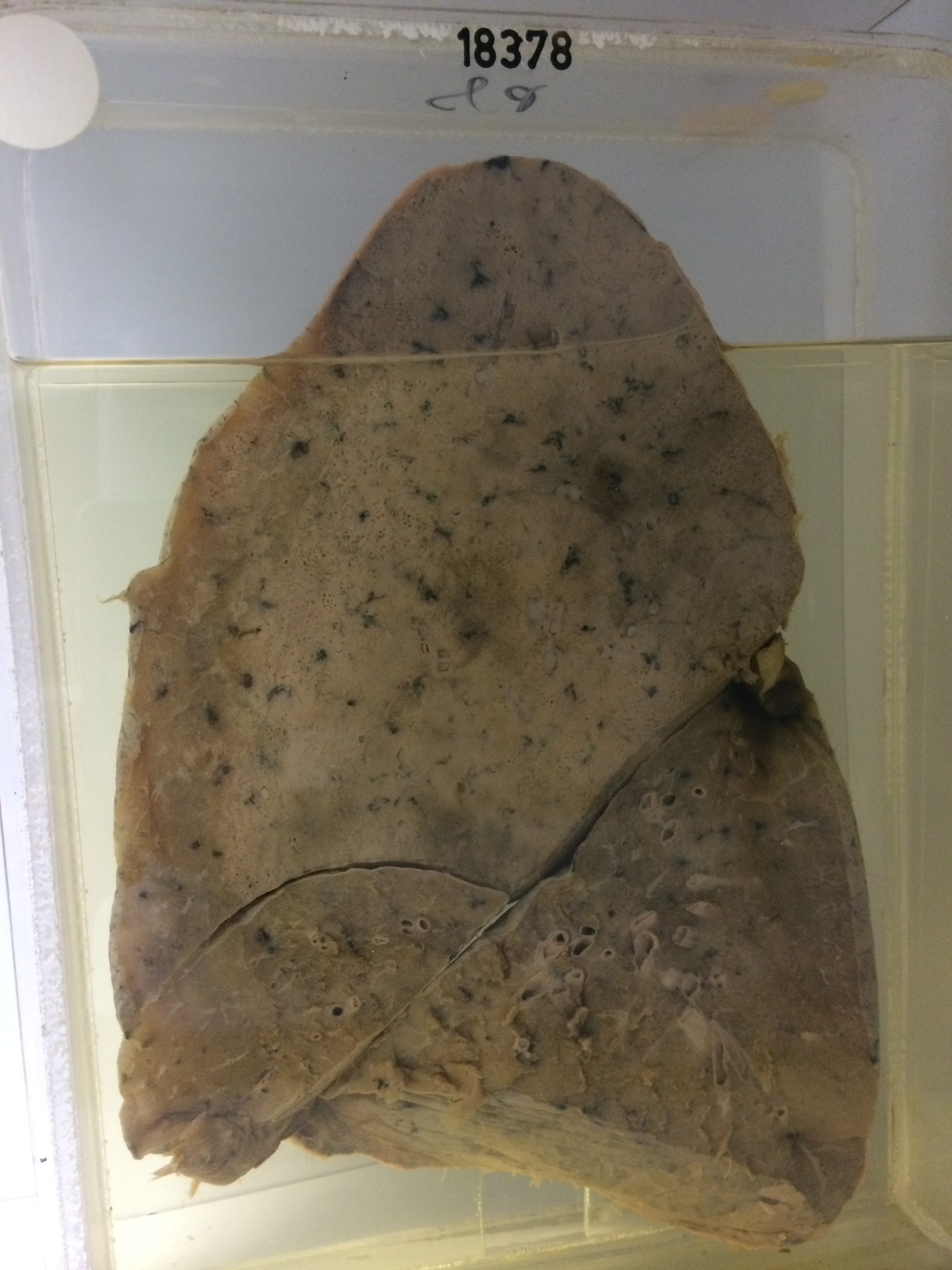
The specimen is of the right lung sectioned to show massive lobar consolidation in the stage of grey hepatisation in the upper lobe, sparing only the lingular segment. Air passages are plugged with exudate. There is marked overlying fibrinous pleurisy. The middle and lower lobes are congested but are not consolidated. Histology shows lobar pneumonia in the stage of grey hepatisation with some small abscesses. Air passages contain mucinous exudate. No culture was taken from the lung.