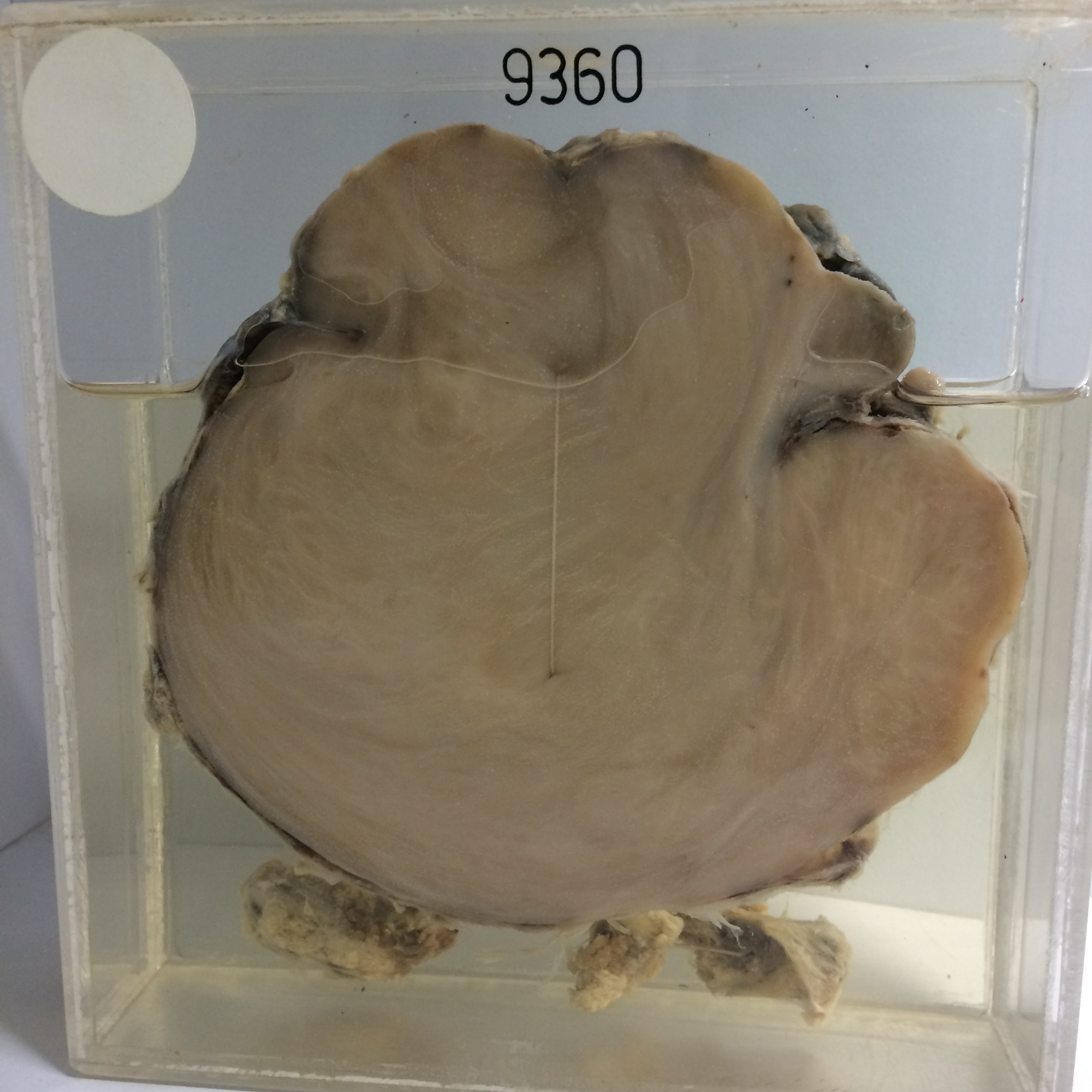
9360 GANGLIONEUROMA OF MEDIASTINUM
A male orchardist aged 22 was found on mass X-ray to have a posterior-superior mediastinal tumour. He had no symptoms. There were multiple pigmented skin moles and there was
X-ray evidence of erosion of a rib and widening of the intervertebral notch of the 4th thoracic nerve on the left side. A pre-operative diagnosis of neurofibroma was therefore made and the tumour was resected in November 1954. At operation the tumour lay outside the pleura but there was an intraspinal prolongation through a greatly enlarged 4th left intervertebral foramen. The tumour was completely removed.
The specimen consists of the tumour and 3 pieces of rib. The largest mass measures 12 cms in diameter and its cut surface shows grey solid whorled myxomatous-appearing neurofibroma. The reverse of the specimen shows the capsule of the tumour formed in part by the parietal pleura. The three fragments of tissue at the bottom of the jar are pieces of rib eroded by the tumour. Histology shows a background resembling a somewhat myxomatous neurofibroma but there are also groups of swollen degenerate ganglion cells.