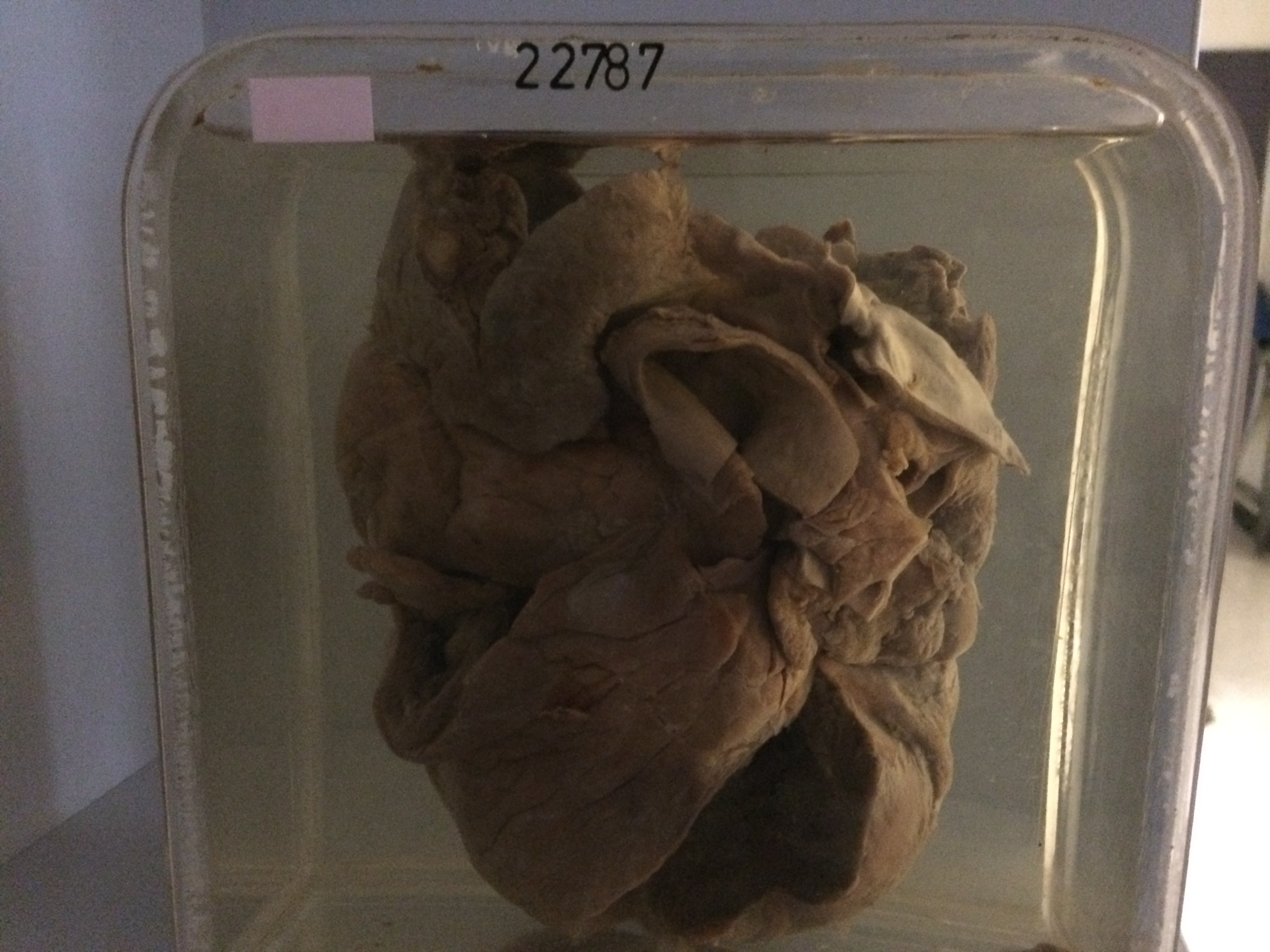
22787 BILATERAL ATRIAL THROMBI
The patient was a man of 70 with a long history of ischaemic heart disease, who had been admitted several times during the previous 12 months with congestive cardiac failure. On his last admission the B.P. was 150/100 and the JVP was elevated to the angle of the jaw. There were bilateral pleural effusions. A good diuresis was achieved but he developed what was thought to be pneumonia in the right lower lobe and died after 2 weeks in hospital. At postmortem there was antemortem thrombus in pulmonary arteries in the right lung with an old breaking down haemorrhagic infarct in the posterior basal segment of the lower lobe, with an overlying encysted fibrinous pleurisy.
The specimen of the heart shows a fibrinous pericarditis. The right atrium is dilated and the tricuspid valve admits 5 fingers. There is a large laminated antemortem thrombus adherent to the antero-lateral wall of the right atrium. The right ventricle is dilated and thin walled. There is antemortem thrombus adherent to the ventricular surface of the tricuspid valve. The left atrium is enlarged and dilated and almost completely filled by a large free antemortem ball thrombus, which has unfortunately been damaged during fixation and mounting. There is also antemortem thrombus in the left atrial appendage. The left ventricle is dilated and thick-walled. There is antemortem thrombus attached both to the septal wall and antero-lateral walls of the ventricle.