22922 HEPATOMA AND CIRRHOSIS
This patient was a woman aged 79 with hypertension, congestive cardiac failure, and dementia resulting from a previous stroke. At her last admission her liver was enlarged and on scan showed large areas of reduced uptake. There was fluctuant jaundice and she died rather suddenly a few days later. At postmortem there was a large amount of fresh blood in the peritoneum and a large haematoma in the lesser sac. It seemed to the prosector that the bleeding point was on the inferior border of the liver where a mass of haemorrhagic and necrotic tumour projected into the peritoneum.
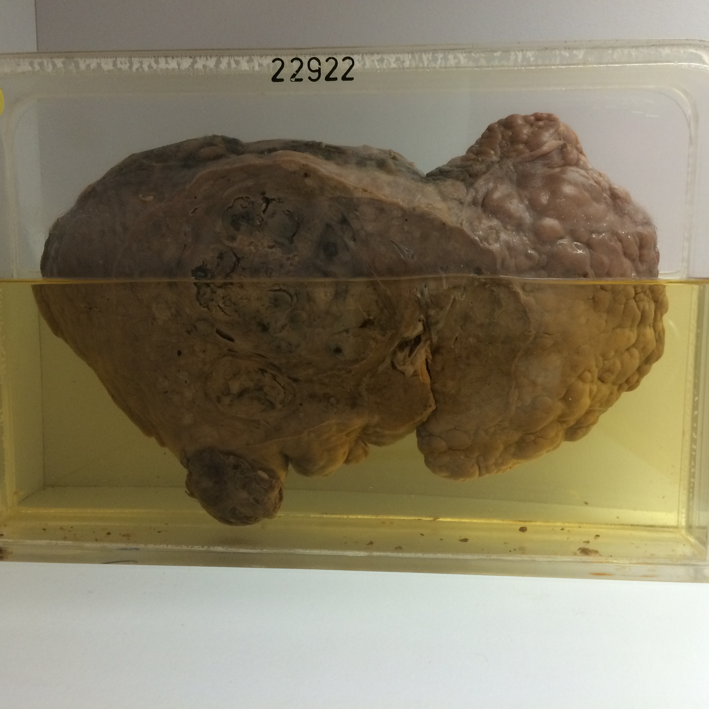
The specimen is the liver displaying gross irregular cirrhosis. Some regeneration nodules are almost 2 cms in diameter. A large hepatoma about 10 cms in diameter is present in the centre of the organ and there are separate nodules of tumour towards the lower border. The cut surface of the tumour is lobular and there are many areas of haemorrhage and necrosis. A haemorrhagic necrotic mass of tumour 3 cms in diameter projects from the lower border of the right lobe.
Histology shows a florid cirrhosis with intense chronic inflammatory infiltration in the fibrous septa and many proliferated bile ducts. The hepatoma consists of comparatively well differentiated tubular and solid-acinar cuboidal-celled structures.