18881 IDIOPATHIC CYSTIC DISEASE, FOCAL EMPHYSEMA AND PNEUMONIA
This 63 year old man had had Parkinson’s disease for many years and then developed thyrotoxicosis with confusion and dementia. Partial thyroidectomy was performed and he was discharged from hospital relatively well. Shortly thereafter his state of consciousness deteriorated and he was thought to have a subdural haematoma, but bilateral burr-holes revealed nothing. Ventriculogram showed symmetrical dilatation of the ventricles and a diagnosis of cerebral atrophy was made. He died from bilateral pneumonia. At postmortem the brain showed slight atrophy of the hemispheres and loss of pigment in the substantial nigra, in which numerous Lewy bodies were present.
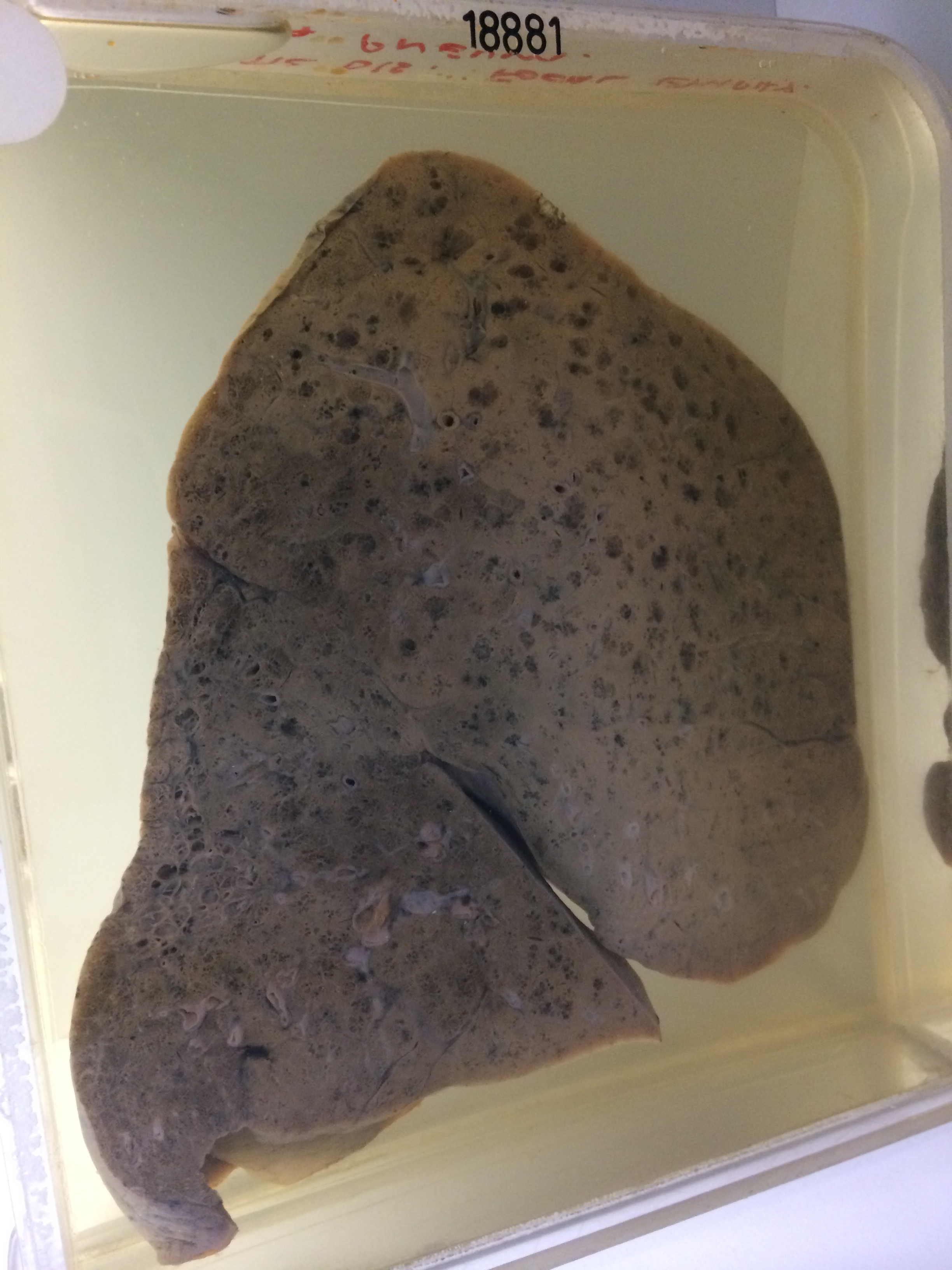
The specimen is the right lung sectioned to show moderately advanced focal centrilobular emphysema maximal in the apical and hilar regions of the upper lobe. Much of the central region of the upper lobe shows diffuse pneumonic consolidation in the stage of grey hepatisation. There is an area of idiopathic cystic disease beneath the pleura of the posterior border of the lung in the apical segment of the lower lobe. Histology shows that the area in the apical segment of the lower lobe consists of large spaces lined by tall columnar epithelium. There is intervening fibrosis and chronic inflammation. Some of the spaces contain pus. The upper lobe shows lobar pneumonia in the stage of grey hepatisation.