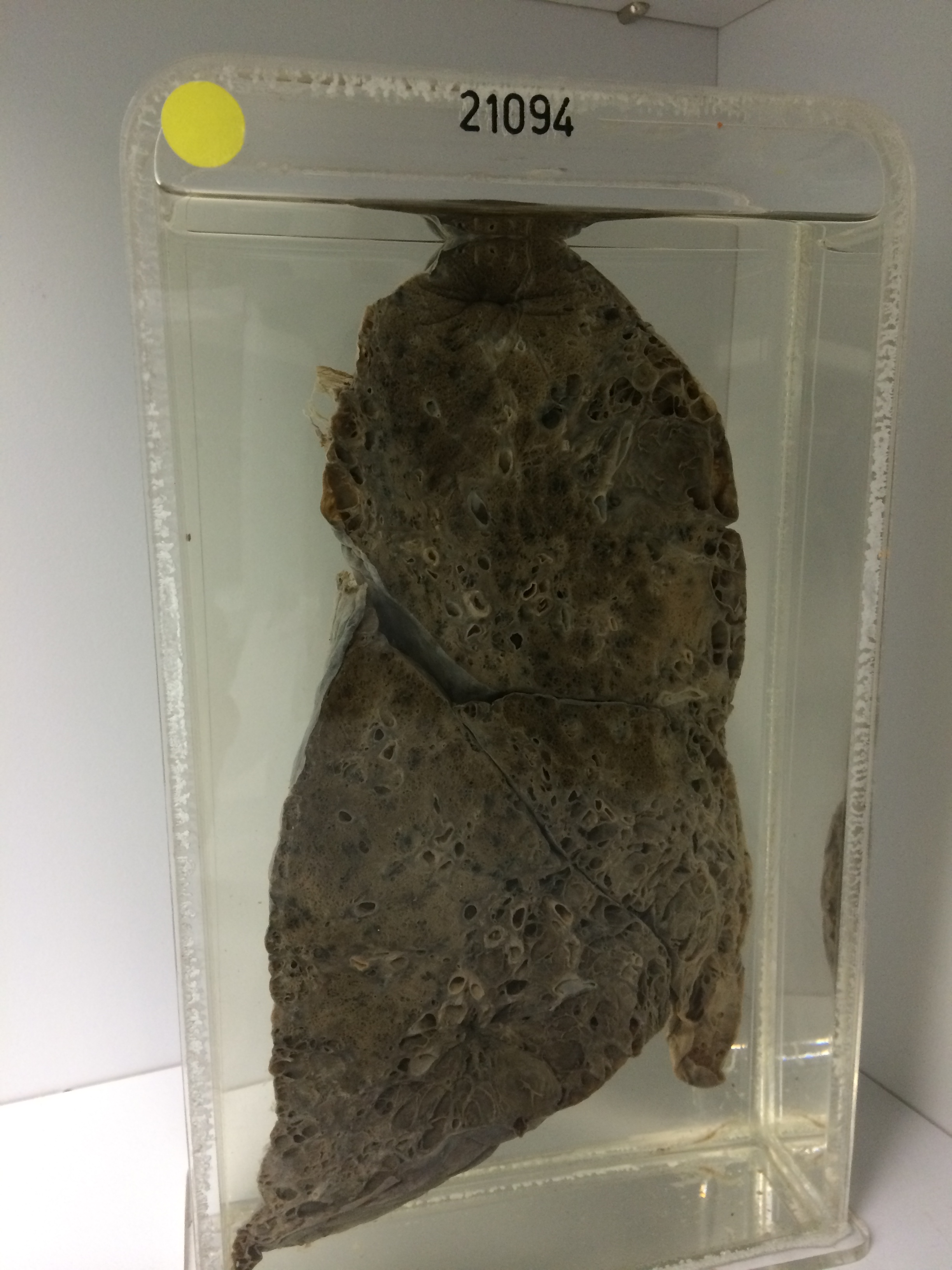
21094 EMPHYSEMA AND FIBROSIS
The patient was a man aged 63 who after extensive investigation was found to have pneumoconiosis. The pulmonary fibrosis was provisionally attributed to wood dust (Meranti) to which he had been exposed for many years. Lung function tests at this stage were said to be not grossly abnormal. On the morning of his last admission he had a sudden attack of coughing with intense dyspnoea, cyanosis and respiratory distress. Examination showed marked finger clubbing, a rapid pulse and an ESR of 118 mms. He died on the day after admission. At postmortem the right ventricle was only slightly hypertrophied and the liver was not congested. Both lungs showed nodular fibrosis and emphysema.
The specimen consists of the right lung sectioned to show large areas of destructive bullous emphysema with spaces measuring up to 1 cm in diameter, crossed by thin spidery fibrous strands. The intervening lung is collapsed and fibrous and there are areas of lipoid pneumonitis. There is some pneumonic consolidation in the lower lobe medial to the cystic areas. Histology shows large cystic spaces lined by bronchial type epithelium which is missing in many places and shows squamous metaplasia in others. The intervening lung is fibrous, collapsed and chronically inflamed. There are areas of lipoid pneumonitis. Some foreign-body giant cells are present. There is haemorrhagic consolidation in the lower lobe.