15143 AORTO-ILIAC GRAFT
The patient was a man aged 83 who presented with a 3-day history of severe constant pain in the left upper quadrant, of sudden onset, which radiated down the left side and to the back, associated with vomiting. On examination the B.P. was 110/70 and there was a large rounded well-defined pulsatile swelling in the left upper quadrant, with overlying tenderness and guarding. There was a soft blowing mitral systolic murmur and a coarse aortic diastolic murmur. A provisional diagnosis of dissecting aneurysm was made. At operation next day a saccular atherosclerotic aneurysm was resected and replaced by a teflon graft. He was anuric for 4 days and then oliguric. The BUN rose to 210 mg% and he died of pneumonia on the 9th day. At postmortem the left kidney was small and there was a large infarct in it. The right kidney was large and there was an infarct at the lower pole. The liver was cirrhotic.
There was thrombosis of the left popliteal artery with gangrene of the left leg and there was venous infarction of the bowel in the superior mesenteric territory.
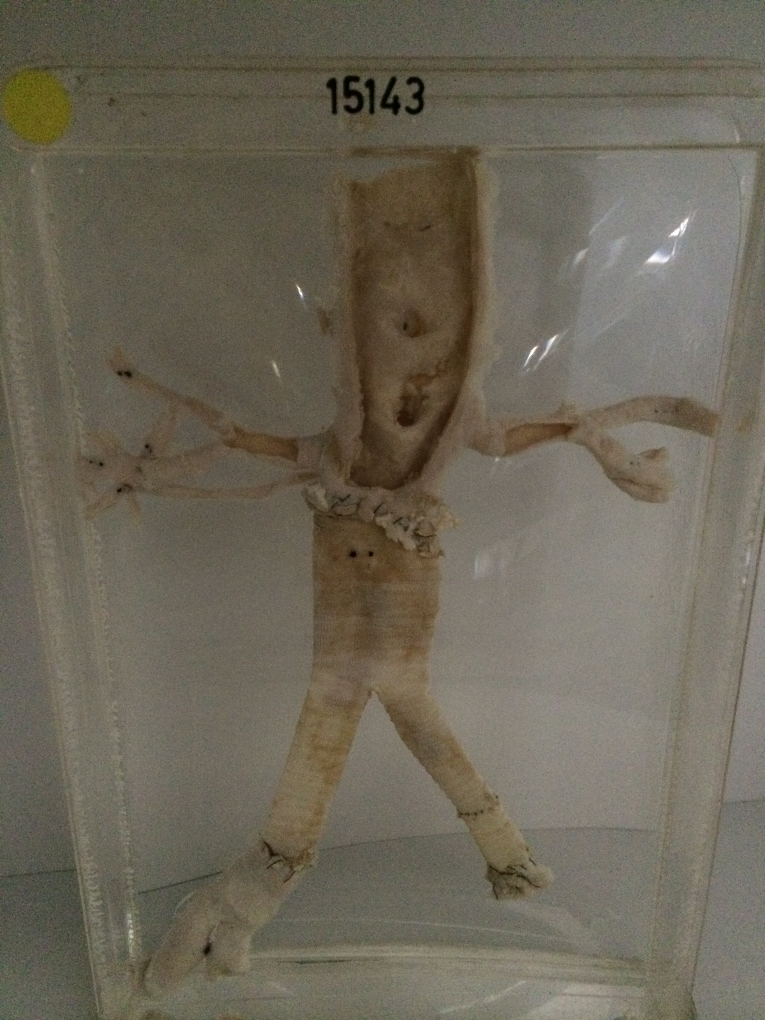
The specimen consists of the lower portion of the abdominal aorta and its major iliac branches viewed from behind. The intima of the aorta around the origins of the coeliac and superior mesenteric arteries shows calcific atherosclerosis but the arteries are not occluded. The main branches of the renal arteries are arteriosclerotic but are free from thrombus. There is an aberrant renal artery on the left side below the main vessel and this was found to be blocked by thrombus at postmortem. The lower 6 cms of the aorta and the proximal 7 cms of the common iliac arteries have been replaced by a teflon graft. The suture lines are intact and there is no thrombus in the proximal ends of the natural arterial channels below the suture lines.