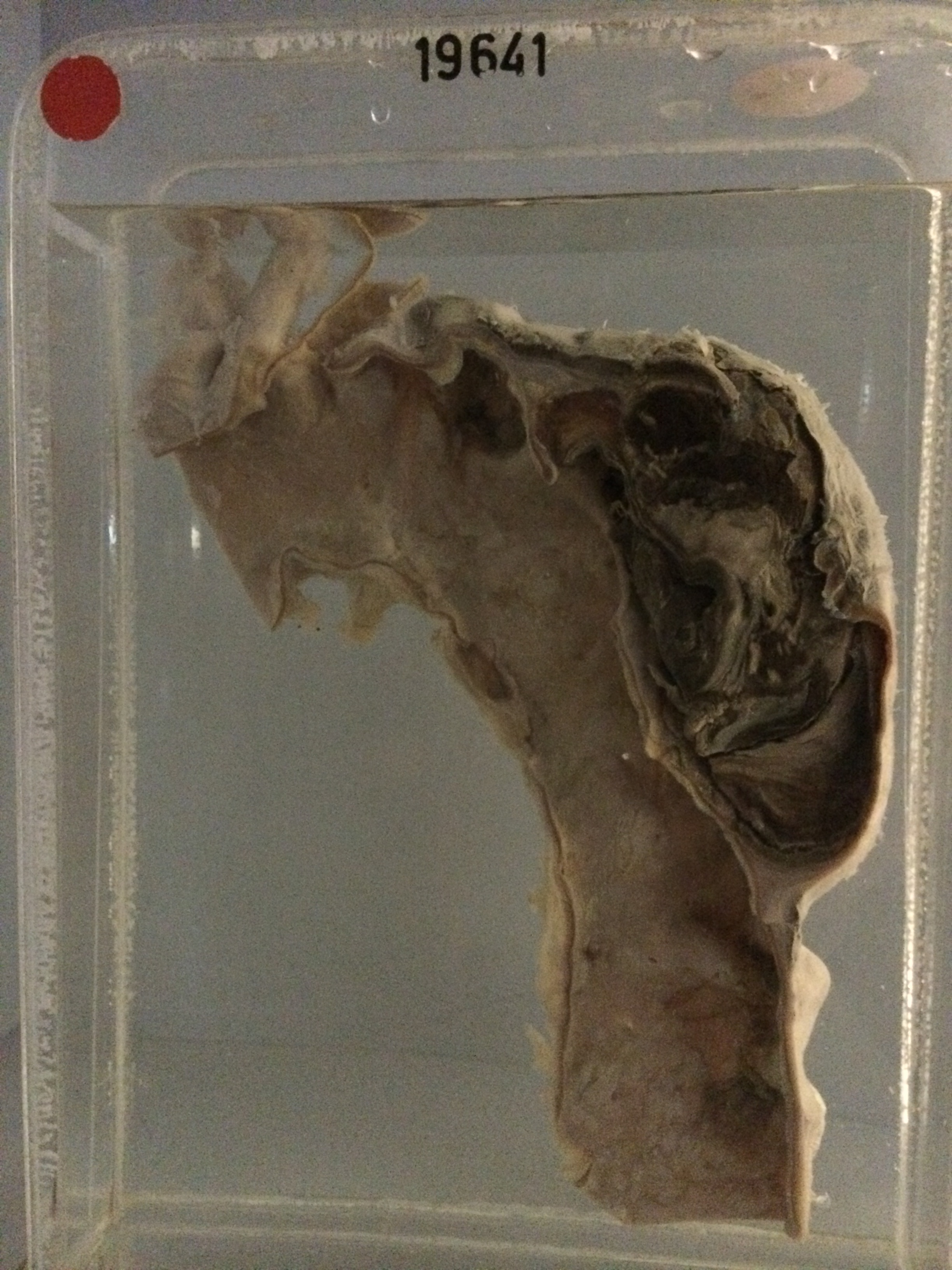
19641 LOCALISED AORTIC ANEURYSM WITH EXTERNAL RUPTURE
This man aged 72 was known to have had a saccular aneurysm of the abdominal aorta for some years. On the morning of admission he experienced a sudden acute severe persistent pain in the left lumbar region and shortly thereafter collapsed. He was diabetic and had had a myocardial infarction 2 years previously. On admission the B.P. was 120/80 and there was a palpable pulsatile mass 10 cms in diameter above and to the left of the umbilicus. The abdomen was opened and a dacron graft was inserted to replace the ruptured abdominal aneurysm. After the operation there was gross oedema of the legs and scrotum and ascites. On the 5th day a delayed rupture of the spleen occurred and the spleen was removed. Two days later there was sudden severe left chest pain and collapse. Paracentesis thoracis produced blood, and a diagnosis of rupture of the thoracic aorta was made. He died next day.
The specimen consists of some 25 cms of the thoracic aorta from the innominate artery to the descending portion. The aorta is generally thickened and there is quite marked pearly intimal fibrosis with some longitudinal and stellate wrinkling. There is a small localised aneurysmal bulge 3 cms in diameter projecting upwards from the arch just distal to the left subclavian artery. Below this aneurysm there is a similar intimal defect 3 cms in diameter with a smooth rolled edge and there is a large haematoma 10 cms long and 5 cms deep. projecting to the left in the substance of the media in relation to this intimal defect. The intramural haematoma is obviously of some duration and consists of laminated thrombus of varying age. External rupture has occurred near the top of the sac. At postmortem the left pleural cavity was filled with 3500 mls of fresh blood. Histology shows longstanding atherosclerosis in the ascending aorta above the lesion, with quite severe loss of elastic laminae in the media and very slight ingrowth of granulation tissue into the outer third of the media. In the descending aorta below the lesion there is marked ingrowth of vascular granulation tissue containing many plasma cells into the outer third of the media. There is a longitudinal medial split filled with fibrous tissue. There is marked pearly intimal fibrosis. Vasa vasorum do not show endarteritis, but this might be a syphilitic aorta.