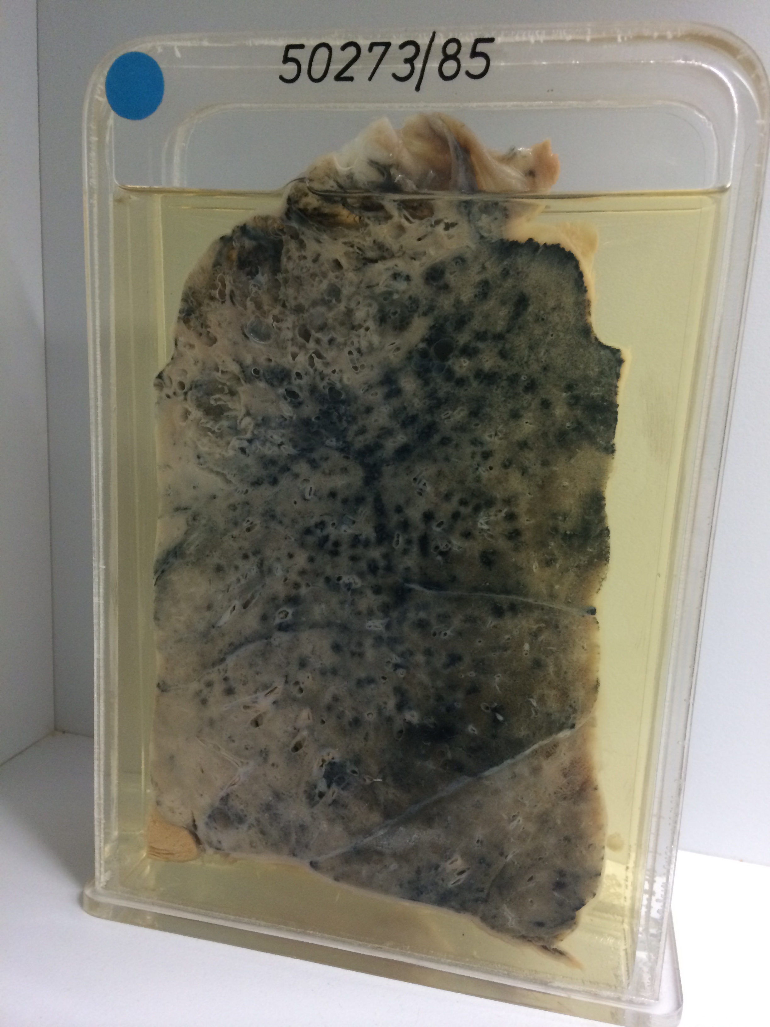
50273/85 PNEUMONIA WITH ABSCESS FORMATION
The specimen shows that the serosal surface of this lung is thickened and has adherent fibrin. At autopsy an empyema was present. The cut surface of the lung parenchyma shows a range of changes. At the apex are some old emphysematous bullae just beneath the pleura. Below this is an honeycombing area of abscess formation. These abscesses are lined by pale necrotic tissue. Very extensive consolidation is present with a wide zone of so-called grey hepatisation. This contrasts with the red lower right-hand portion. These zones illustrate progression of the pneumonic process. The red congested lung changes to grey when all of the alveoli are filled with leukocytes. In this case resolution has not occurred and the disease has progressed to abscess formation. The patient was known to have Legionella pneumonia, but at autopsy Pseudomonas aeruginosa was grown from the lung.