50493/83 HYPERTROPHIED LEFT KIDNEY WITH CORTICA ADENOMA
The patient was a woman aged 61.
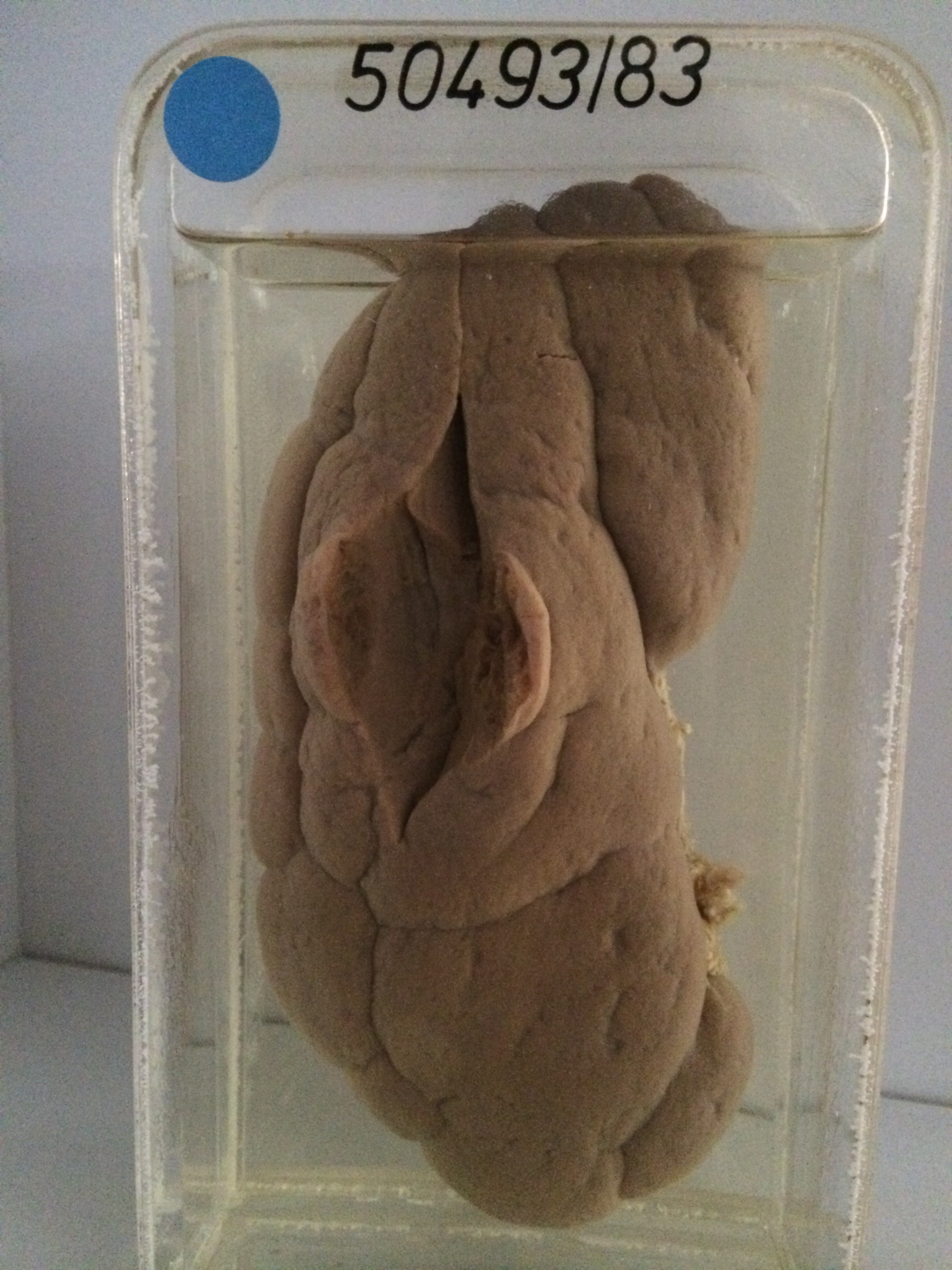
The specimen consists of a very large kidney weighing 300 gms. The cut surface is quite normal with clearly defined cortex, medulla, pyramids, calyces and pelvis. Hypertrophy has occurred because the right kidney was removed surgically at some time in the past. The capsule has been stripped off the surface of the kidney which shows persistence of foetal lobulation. A 25 mm cortical adenoma is present and has been sliced open to show its variegated and partly cystic substance.
Adenomas are similar histologically to renal cortical carcinomas and the distinction is based mainly on size. Tumours under 25 mms do not usually metastasise and are called adenomas