22026 SECONDARY CARCINOMA AND FOCAL EMPHYSEMA
This patient was a man aged 60. Six months before his death he suffered a subarachnoid haemorrhage and an aneurism on the left middle cerebral artery was clipped. Four months later he presented again with pain in the knee. X-ray showed a translucent area, probably a secondary deposit, and chest X-ray then showed multiple metastases throughout both lung fields. He continued to complain of pain in the right knee and died undiagnosed after 6 weeks in hospital. At postmortem there were multiple neoplastic deposits in both lungs, the head of the pancreas, many lymph nodes, the right tibia, both kidneys, lower, small bowel and caecum.
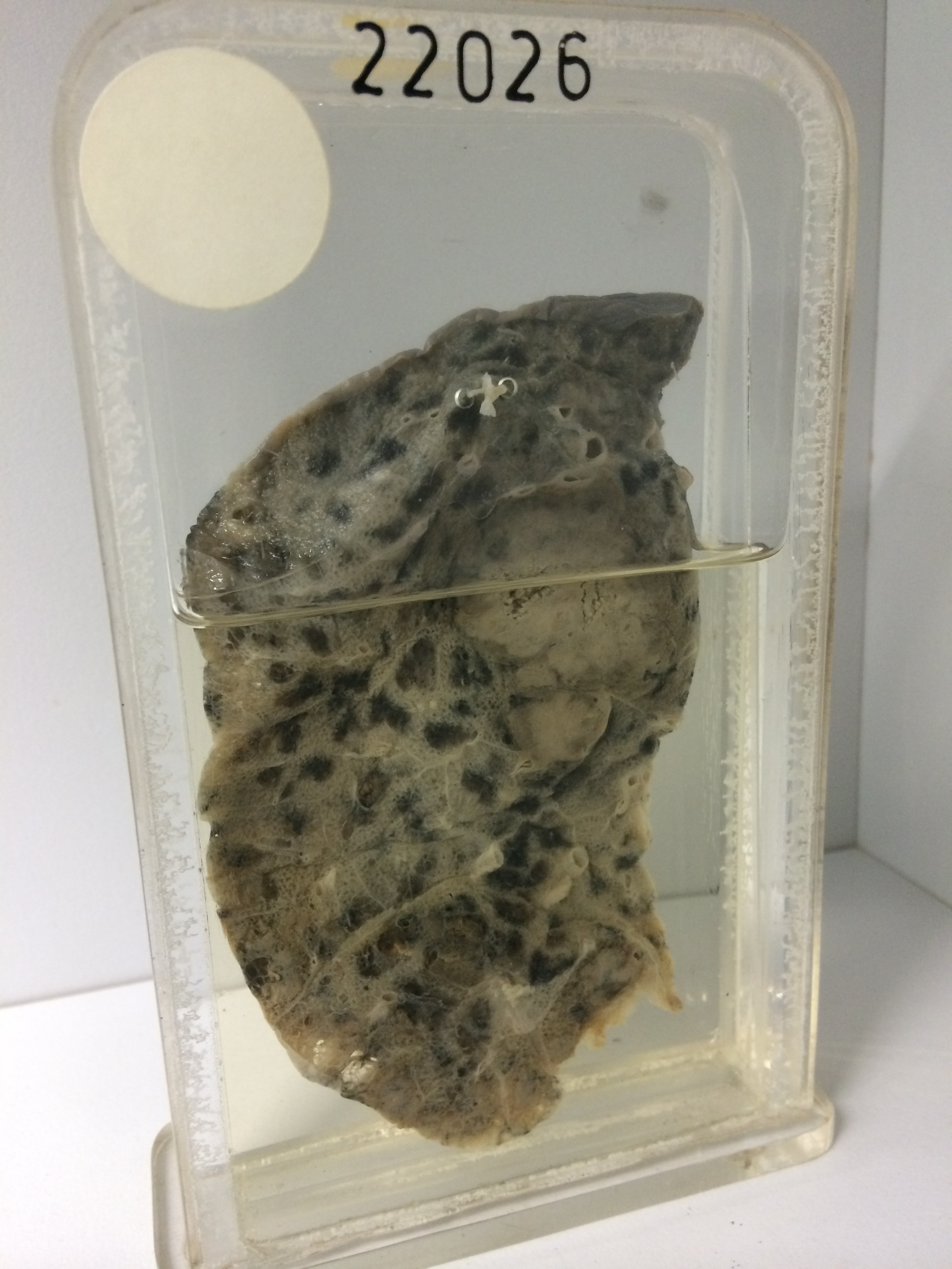
The specimen is a slice of lung in which there are two rounded secondary deposits, the larger measuring 3.5 cms in diameter. Their cut surfaces are brown, soft and cellular and they contain pigment which has been driven to the periphery of the tumour. The remainder of the lung shows advanced focal centrilobular emphysema with anthracosis. The site of the primary is uncertain. The tumour masses are largely necrotic and haemorrhagic. Surviving tumour cells are large, with water-clear cytoplasm and dense hyperchromatic nuclei. Mitotic figures are numerous. The tumour is probably of epithelial rather than connective tissue origin.