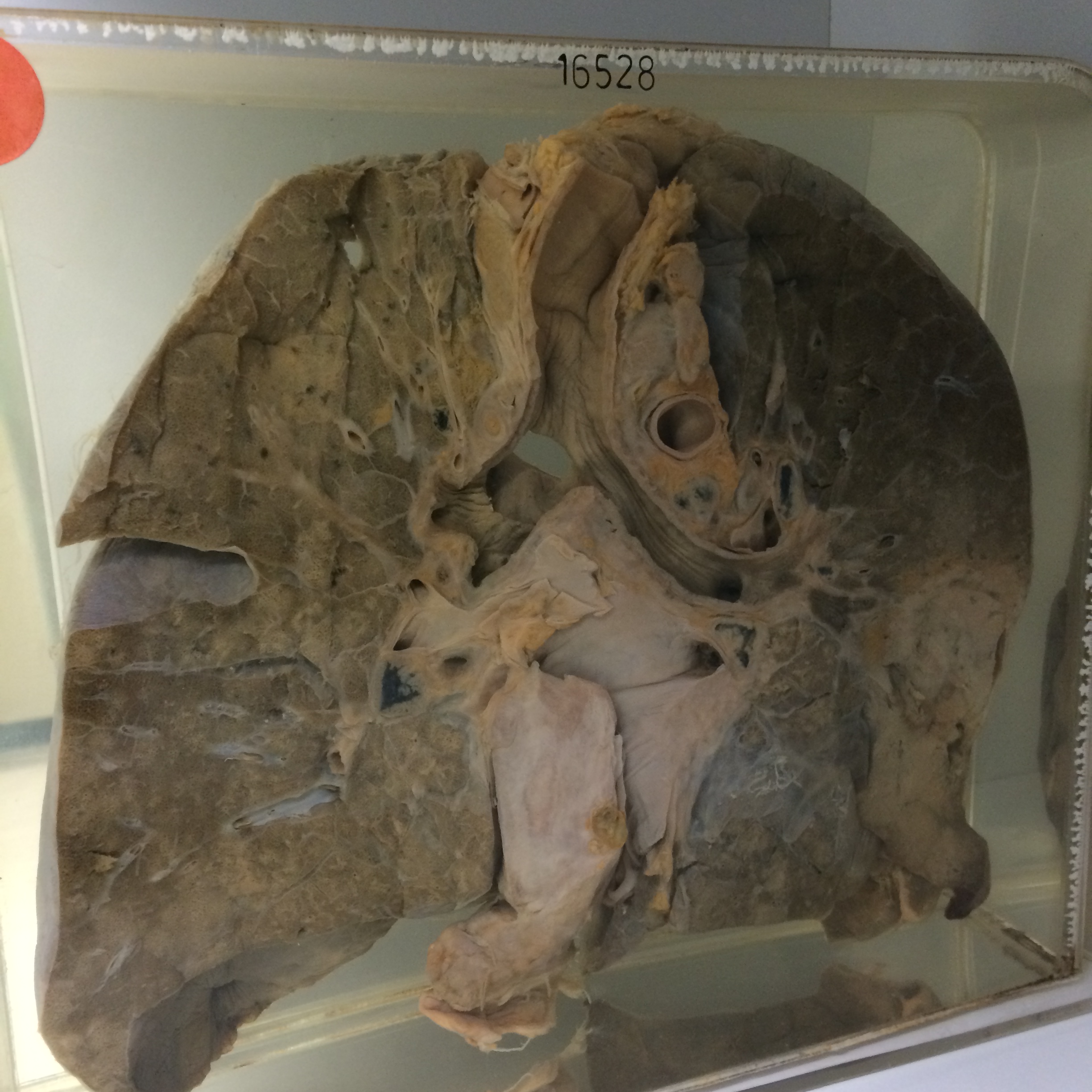
16528 HODGKIN’S DISEASE
This 42 year old man had Hodgkin’s disease for 10 years treated by intermittent courses of nitrogen mustards, cyclophosphamide and radiotherapy. A month before his death he presented with dysphagia, and a barium swallow revealed oesophageal indentation by enlarged mediastinal glands. A tracheo-oesophageal fistula developed with consequent pulmonary infection and he died shortly thereafter.
The specimen consists of the lungs and air passages sectioned to show a massive fistulous opening extending between the origin of the left main bronchus and the oesophagus. The opening of the fistula is about 2 cms in diameter and the major air passages are acutely inflamed. In the right lower lobe is an area of opaque consolidation probably the result of lymphomatous infiltration. Lymph nodes in the mediastinum are involved and there is involvement of the parietal pericardium. Acute bronchopneumonic patches are visible in the left lower lobe. The reverse of the specimen shows a very large lymphomatous mass 6 cms in diameter in the apex of the lower lobe and bronchopneumonic patches in the congested lung beneath it. Histology shows pleomorphic Hodgkin’s disease poor in lymphocytes and rich in fibrous tissue. Reed-Sternberg cells are present.