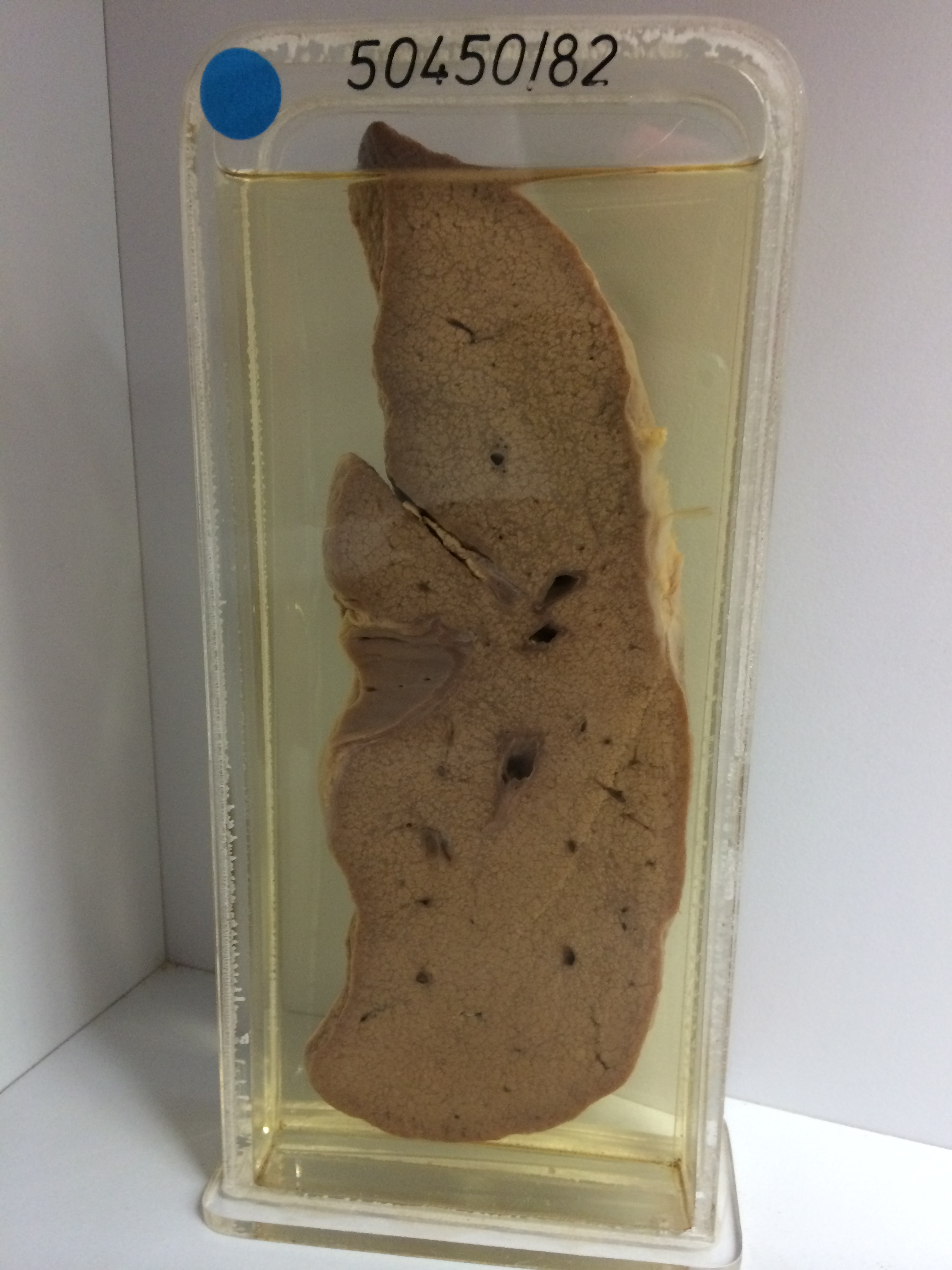
50450/82 MICRONODULAR CIRRHOSIS
The specimen consists of a slice of liver. The cut surface is finely nodular. The nodules measure between 1 mm and 2 mm in diameter. The nodules are surrounded by pale grey fibrous tissue. The parenchymal changes are reflected by fine nodularity of the serosal surface. A few vessels contain blood clot. This is postmortem clot, not antemortem thrombus. This can be stated with some certainty as there is no congestion of the liver evident. The nodules are pale because there is also steatosis present. In steatosis the liver cells are distorted by droplets of fat within the cytoplasm.
Last modified: Wednesday, 2 August 2017, 8:57 AM