22833 MASSIVE PULMONARY INFARCT AND TUBERCULOUS PNEUMONIA
This 50 year old man was a private patient admitted to a local hospital with a 2-day history of nagging cough, breathlessness and haemoptysis. Three weeks before this he had had an influenza-like illness but recovered without incident in a week. Chest X-ray after admission showed right lower lobe consolidation. Swinging fever up to 39oC developed on the 2nd day and persisted until death. On the second day he developed superficial thrombophlebitis and he continued to produce large amounts of purulent bloodstained sputum. Ampicillin was begun, together with intravenous heparin and other autocoagulants. Between the 3rd and 5th week his general condition improved but X-ray showed further patchy pulmonary opacities which the radiologist thought were due to fresh pulmonary emboli. The inferior vena cava was the ligated but he died shortly after the operation. At postmortem there were scattered rounded yellow consolidated areas in the left lung.
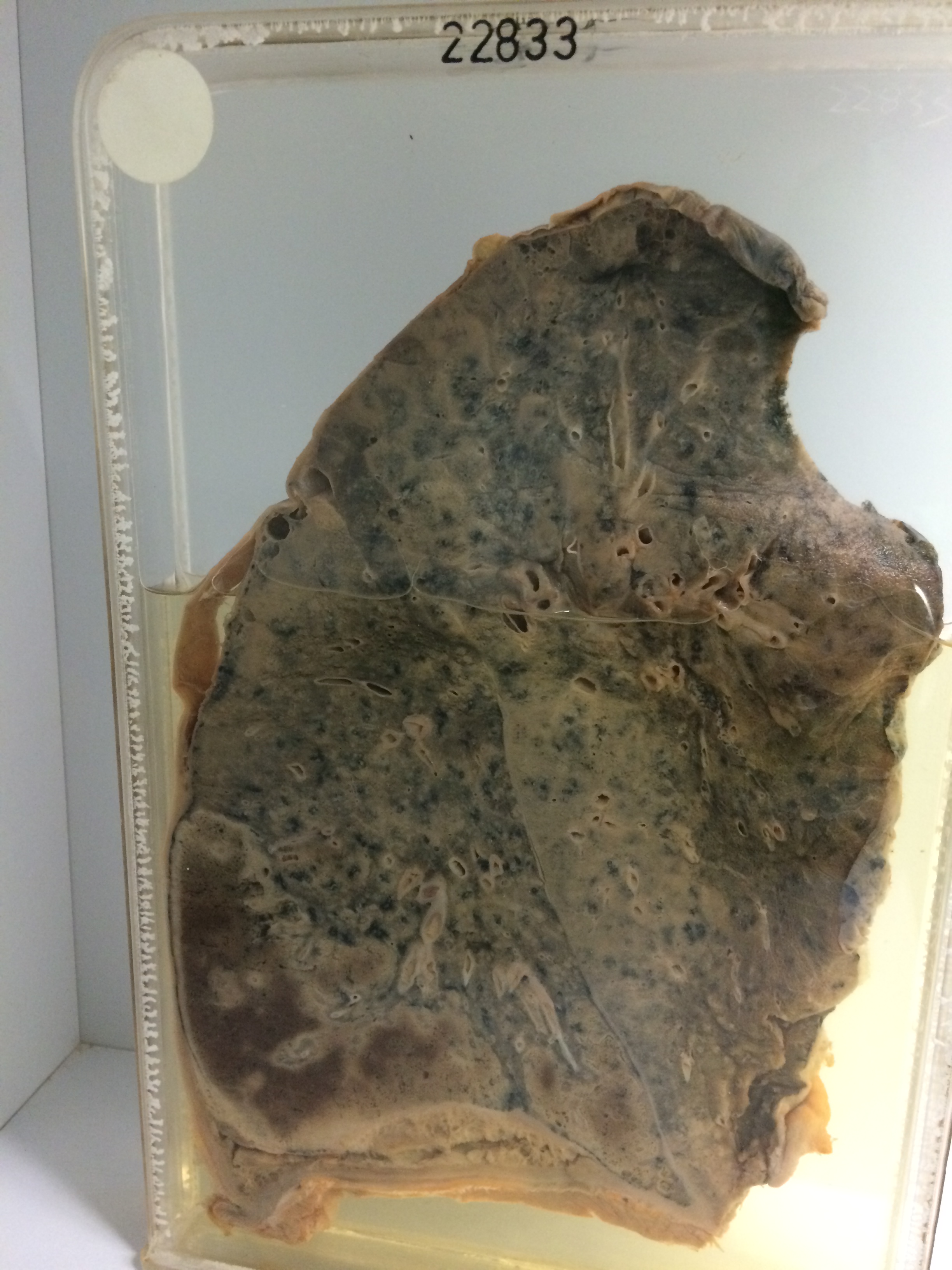
The specimen is of the right lung. There is massive haemorrhagic and ischaemic pulmonary infarction of some duration in the posterior basal and anterior basal segments of the lower lobe. There is marked overlying fibrinous pleurisy with adhesion of the lung to the parietal layer. Masses of antemortem thrombus are visible in the arteries to the infarcted area. The lower lobe above the infarct shows scattered yellow-grey opaque bronchopneumonic rosettes. In the posterior border of the upper lobe there is diffuse pneumonic consolidation, which extends as a continuous area into the lingular segment along the interlobar fissure. Histology shows confluent caseous foci with typical giant cells, and spreading tuberculous pneumonia with plentiful fibrin and epithelioid cells.