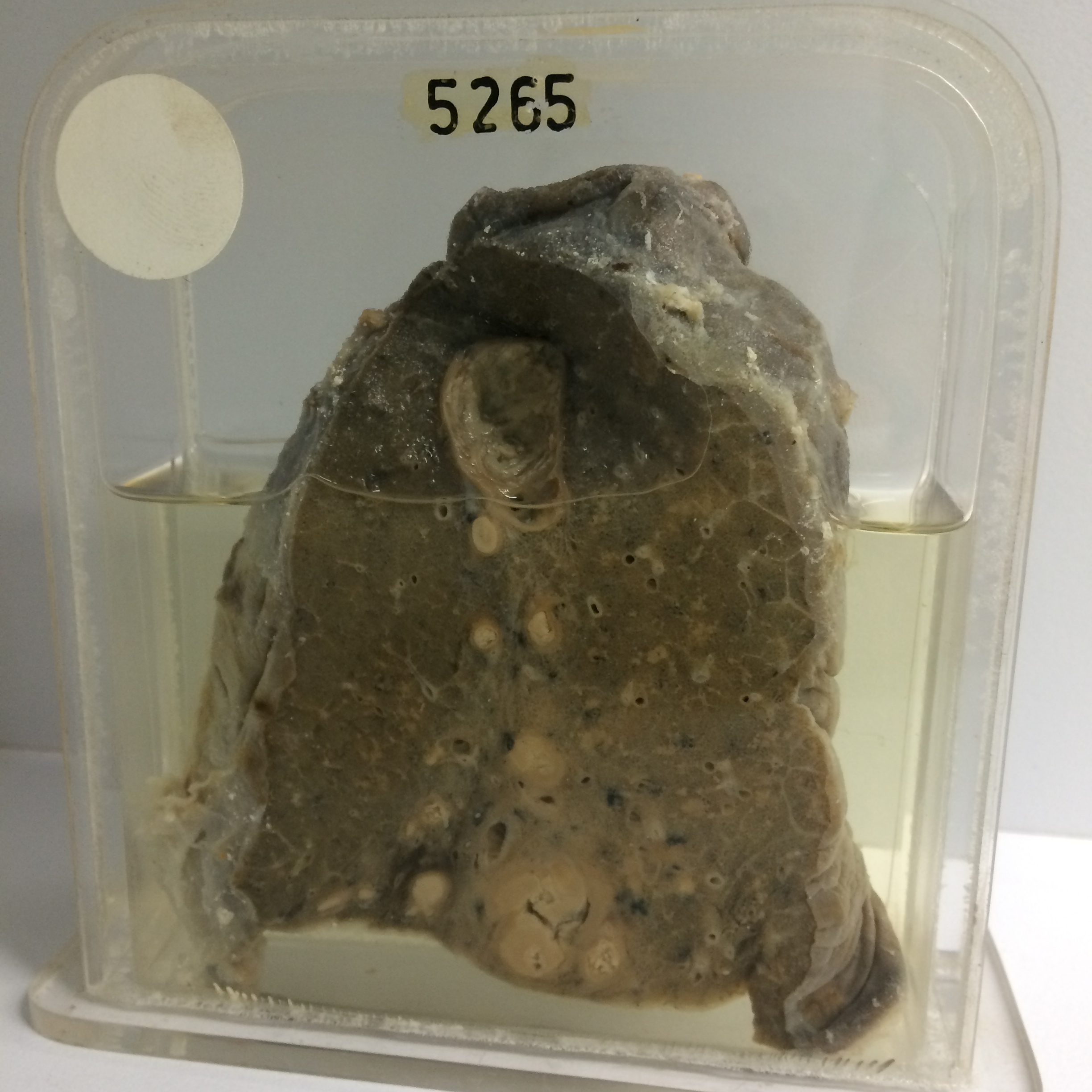
5265 CHRONIC FIBRO-CASEOUS TUBERCULOSIS
The patient was a man aged 23 who had migrated to Australia from Europe 2 years previously. Chest X-ray on arrival was normal. A year later he developed a cough with sputum. X-ray in a country hospital then showed infiltration at the left base thought to be tuberculous. On admission to the R.A.H. a few weeks later there was left basal cavitation and the sputum was positive for tubercle bacilli. He was treated by streptomycin. Eighteen months later the sputum was still positive but the pulmonary infiltration was a little smaller. The lower lobe was then resected.
The specimen consists of this lobe cut to show several chronic caseous tuberculous lesions. The largest is a cavity in the apical segment and measures about 2 x 1.5 cms. Below this are small rounded inspissated caseous lesions with dense fibrous encapsulation. A group of encapsulated caseous lesions are present at the base. Small acute diffuse miliary foci are visible in the intervening lung tissue. Histology confirms that the small scattered foci are miliary tubercles. There is unfortunately no record of the patient’s subsequent fate.