23026 ISCHAEMIC COLITIS
This is a surgical specimen from a woman aged 76. The symptoms begin with severe central abdominal pain, followed by many loose bowel actions with relief of the pain. There was some nausea but no vomiting. During the latter part of the diarrhoea she passed a good deal of dark blood per rectum. There was mild general abdominal tenderness but rectal examination showed no abnormality. A barium enema was then performed, which showed the typical ‘thumb printing’ appearance of ischaemic colitis extending from the middle of the transverse colon and around the splenic flexure to about the proximal third of the descending colon. She was first treated conservatively with drip and suction, but after 9 days of gradual deterioration the affected portion of bowel and resected.
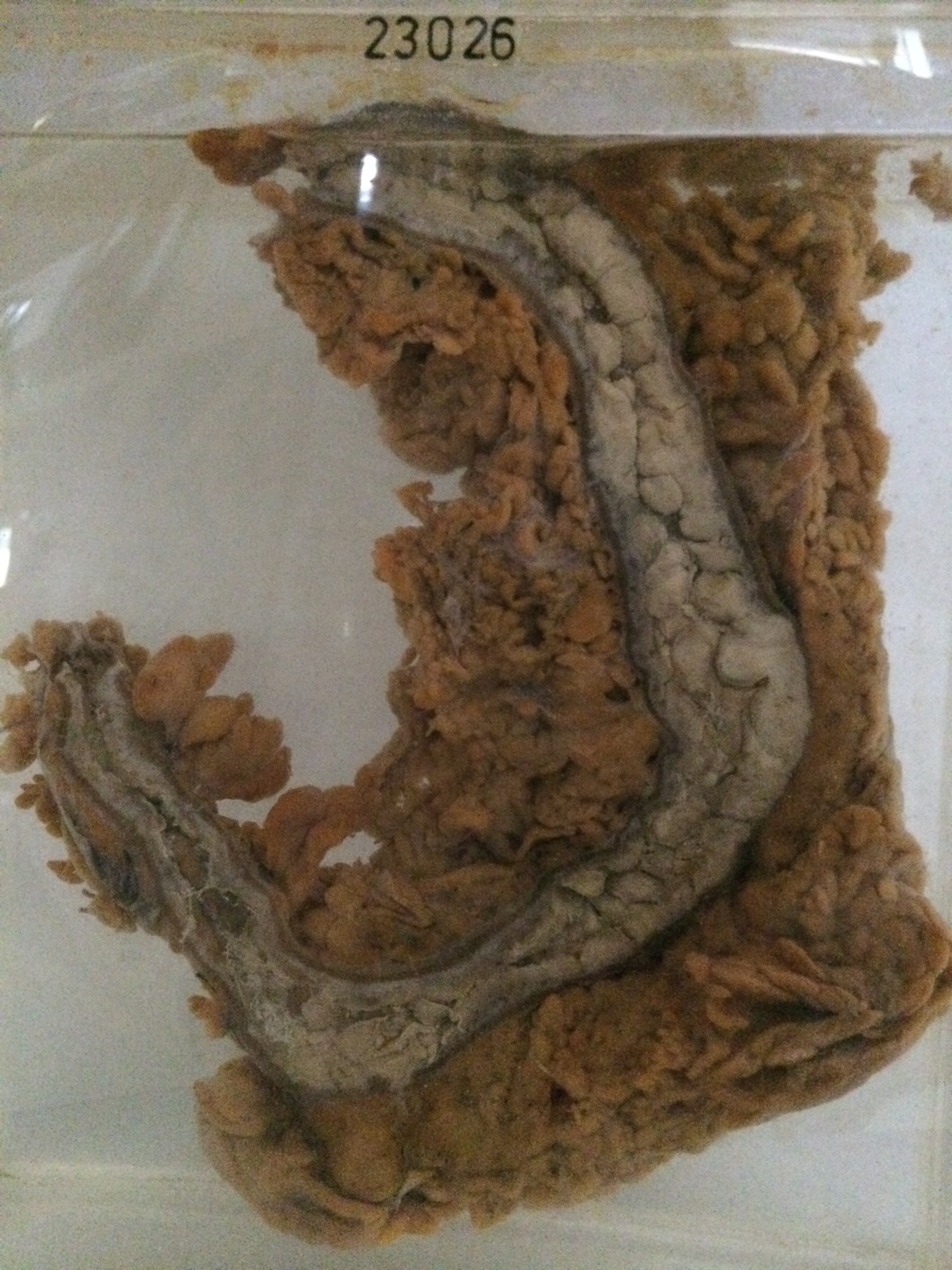
The specimen consists of some 30 cms of colon together with attached omentum. The omentum is thickened and adherent to the colon in several places. There is gross thickening of the mucosa which pale and necrotic and raised into many nodular elevations about ½ to 1 cm in diameter. The muscle coat is thickened. Histology shows swelling and necrosis of the mucosa with superadded infection and much friable exudate. There is fibrous granulation tissue in the deeper parts of the submucosa and there is some inflammatory oedema and congestion between the thickened muscle bundles. In one place there is an incipient perforation, with only a few strands of serosa and subserosa remaining viable.