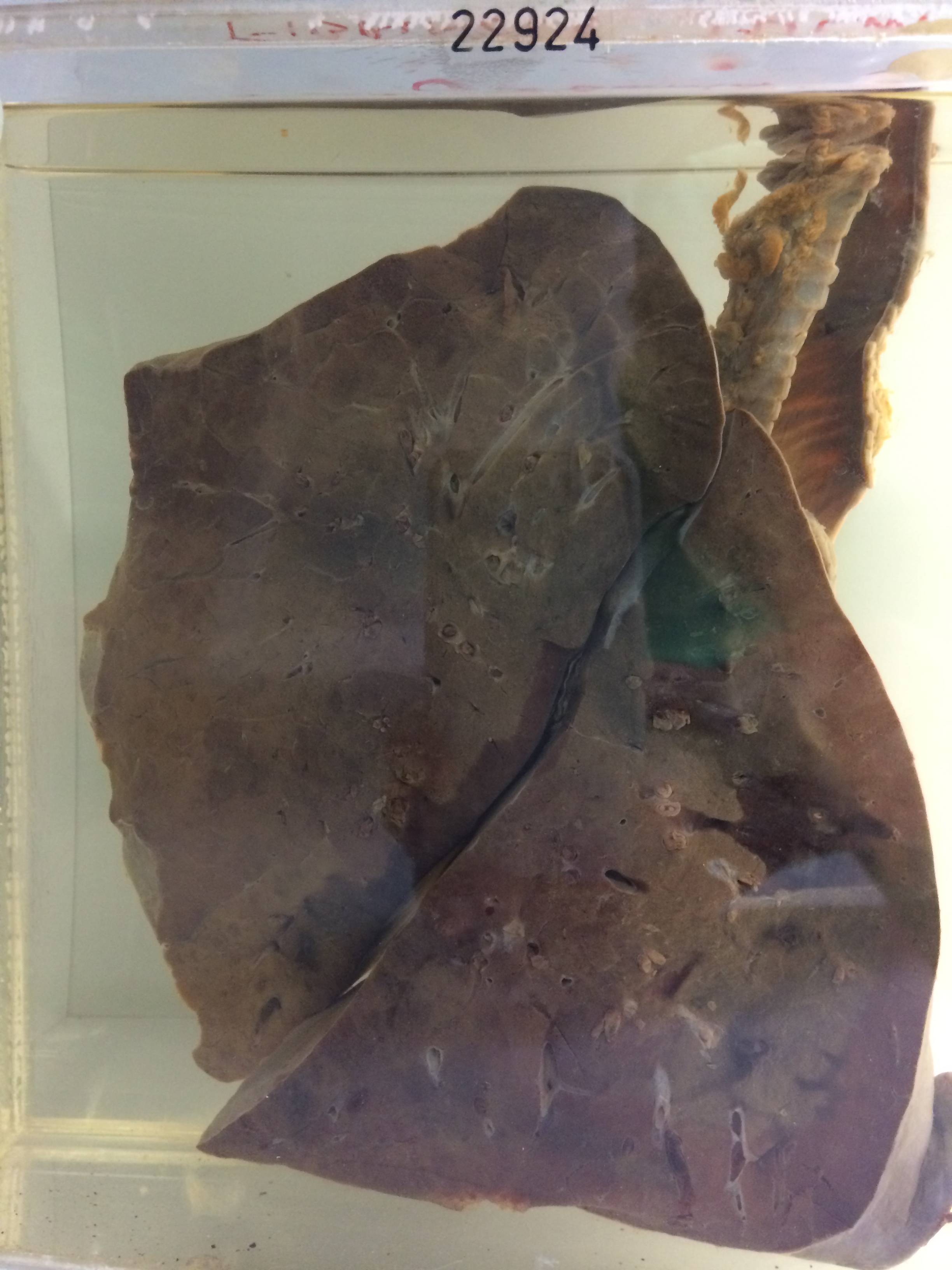
22924 FIBRINOUS PULMONARY OEDEMA
The patient was admitted to hospital after being involved in a vehicular accident. He was concussed and had evidence of fractured ribs and scapula with a ruptured intra-abdominal viscus. A laparotomy was performed and the ruptured right kidney removed. He subsequently developed respiratory and renal failure. Despite intensive treatment, including dialysis, his condition steadily deteriorated and he died about one month after the accident.
The specimen consists of a portion of the left lung showing intense generalised haemorrhagic consolidation involving almost the entire lung except for a rather narrow strip subpleurally anteriorly which is only patchily affected. An antemortem thrombus is present in the left main bronchus, completely obstructing the lumen. Histology shows congestion of alveolar walls and interstitial swelling in some places. There is a dense patchy fibrinous exudate containing many mononuclear cells but few polymorphs. Widespread loci of early fibroblastic organisation are evident in the exudate. Small vessels are congested and are packed with polymorphs. There are numerous small focal haemorrhages, especially in the lower lobe.
(Comment:- This condition was first recognised radiologically and was originally described as ‘uremic lung’. However it occurs in other conditions characterised by left ventricular failure such as essential hypertension and acute rheumatic fever. Some degree of chronicity is necessary as it does not occur in acute left ventricular failure due to myocardial infarction.
In addition to the left ventricular failure there appears to be a capillary toxin involved. The initial change is a fibrin-rich pulmonary oedema which later progresses to intra-alveolar and intra-ductal fibrosis. There is some evidence for impaired fibrinolytic activity as well).