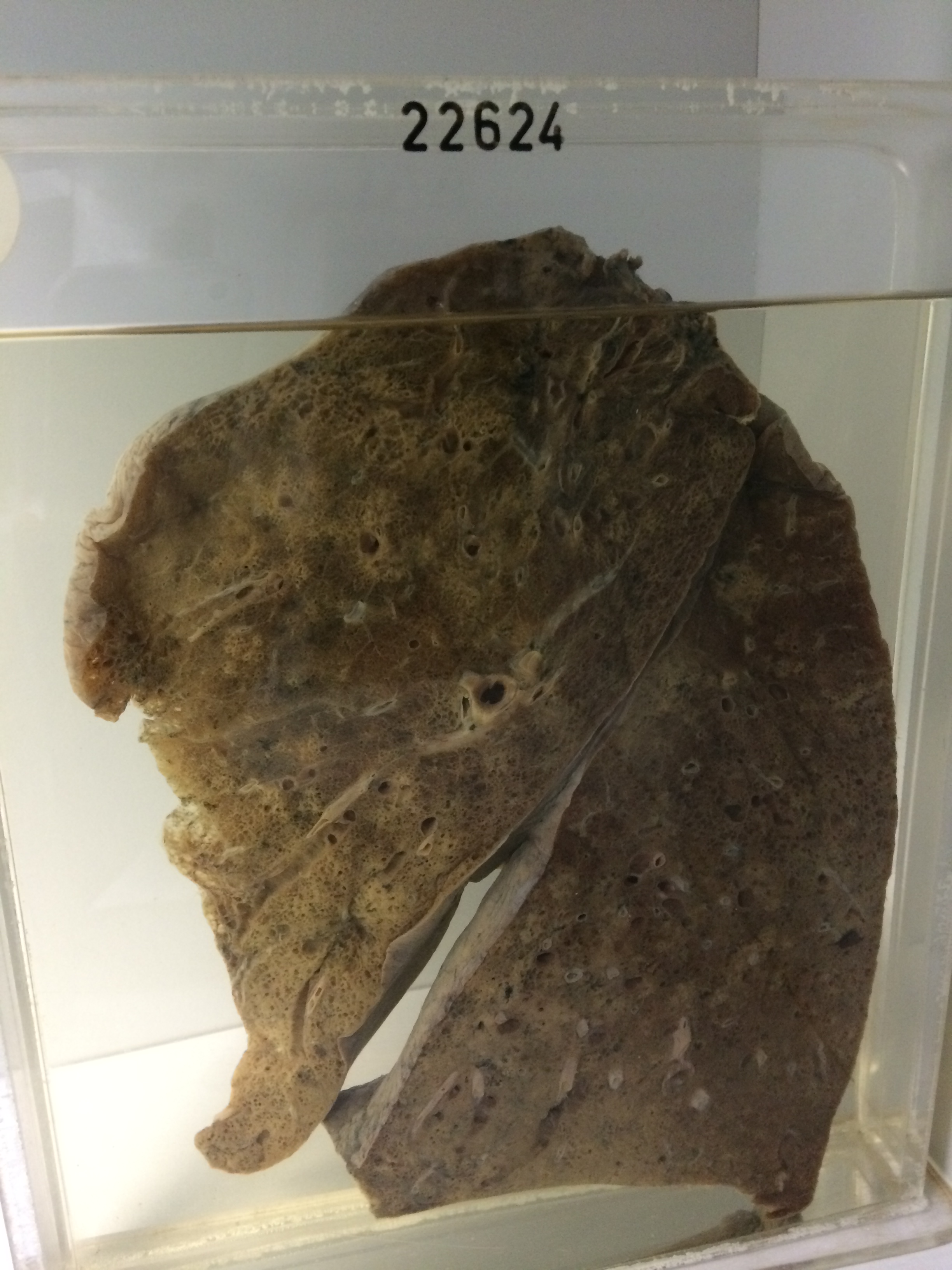
22624 CHRONIC SUBPLEURAL CYSTIC FIBROSIS & SUPPURATIVE BRONCHOPNEUMONIA
The patient was a man aged 84 who was admitted with left basal pneumonia. In hospital he became cyanosed and X-ray showed mottled opacities throughout both lungs. There was a leucocytosis of 13,600 and the ESR was 107. Sputum culture was negative. He died on the 8th day.
The specimen consists of the left lung showing apical scarring with subjacent emphysema, and patches of bullous emphysema on the anterior subpleural border of the lingular segment of the upper lobe. In addition there are scattered pale patches of bronchopneumonic consolidation throughout the substance of the lower lobe. Much of the lingular lobe shows widespread cystic fibrosis with cysts varying up to 3 mms in diameter. The left lower lobe shows a large posterior patch of subpleural cystic fibrosis with a serpiginous inner boundary between it and the internal aspects of the lobe. Similar changes are present beneath the pleura on the diaphragmatic surface. The substance of the lobe shows widespread rosettes of bronchopneumonic consolidation, going on to the formation of small abscesses in places.