6772 INTRALOBAR SEQUESTRATION
The patient was a woman aged 20 who presented with a febrile illness associated with pus in the left chest. This was diagnosed as an empyema and drainage was performed under local anaesthesia. At the operation it was apparent that the collection of pus was in the lung substance and not in the pleura, and that the condition was in fact an infected congenital cyst of the lung. After drainage for some time a left pneumonectomy was performed. At the operation three large abnormal arteries were found to arise from the aorta to supply the cystic abnormality in the lung, showing that the condition was an intralobar sequestration.
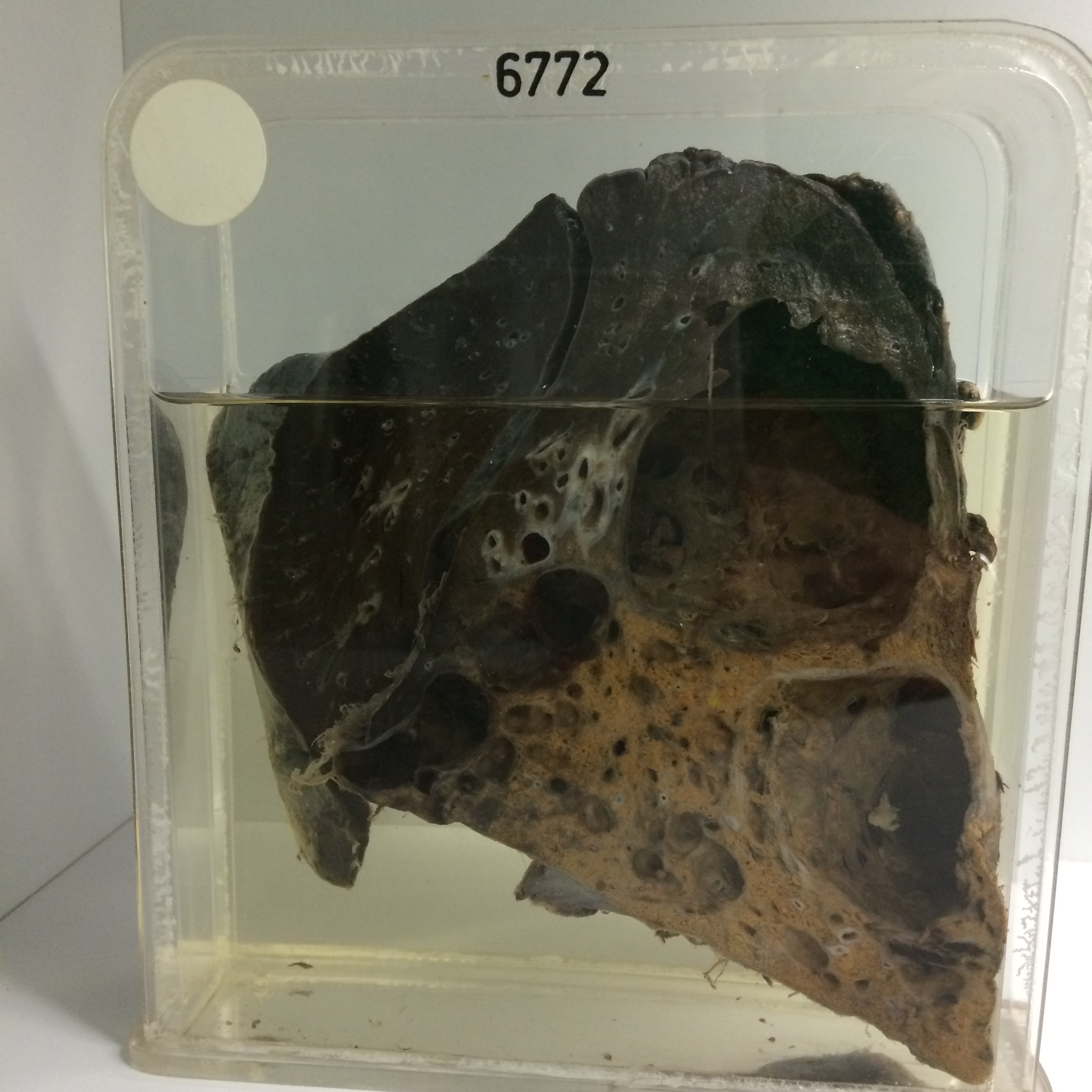
The specimen consists of the sectioned lung. Much of the lower lobe is replaced by a collection of thick-walled cystic cavities. The largest in the apical segment measures 6 cms in diameter. The cysts have intensely congested walls to which purulent exudate adheres. The intervening lung substance in the basal region shows marked lipoid pneumonitis. The overlying pleura is thickened. The three abnormal arteries are visible entering the posterior segment on the medial side. Histology shows large cystic spaces lined by tall columnar epithelium and packed with foamy macrophages. Here and there are cholesterol crystals surrounded by foreign body giant cells. Round cells, foamy macrophages and lymphoid follicles are quite numerous in the interstitial tissues.