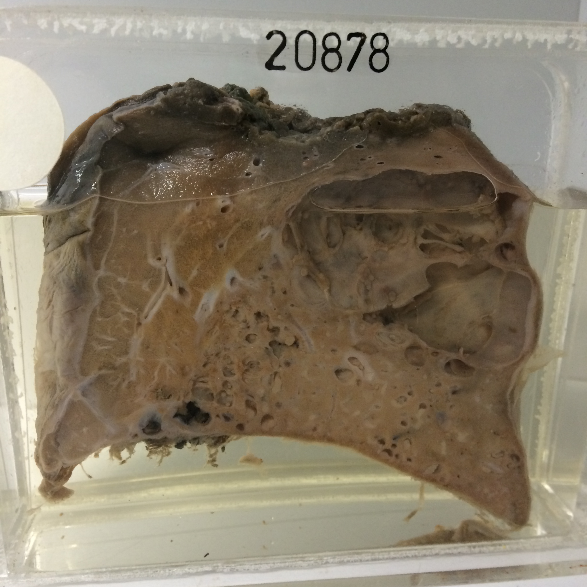
20878 INTRALOBAR SEQUESTRATION
This patient was a boy of 14 with a history of pneumonia 3 months previously, with loss of weight and a productive cough. The affected lobe was resected.
The specimen consists of a thick slice of this lobe. A large cavity consisting of two intercommunicating cysts which are together 6 cms in diameter lies just beneath the pleura. The lining of the cyst is mainly smooth but there are some areas in which pale exudate adheres to the surface. The remainder of the lung shows groups of small cysts with fibrosis and lipoid pneumonitis in the intervening lung tissue which is generally airless. The reverse of the specimen shows in the centre of the affected area a greatly dilated abnormal artery 5 mms in diameter. Histology shows fibrosis and cystic cavities of varying size lined by bronchial type epithelium. There are areas of lipoid pneumonitis in which fat-filled macrophages are prominent. The abnormal artery has thick elastic walls.