21353 CIRRHOSIS WITH HEPATOMA
The patient was a Polish migrant aged 66. He had had chronic bronchitis for 42 years. There had been anorexia and epigastric fullness for 3 months, together with pain in the left subcostal region unrelated to meals. On examination the fingers and toes were clubbed, there was ascites, the liver was enlarged 10 cms beneath the right costal margin and its edge was irregular and tender. The spleen was palpable 5 cms below the left costal margin. There were spider naevi on the chest, a microcytic anaemia, the blood platelets numbered 80,000 and prothrombin activity was 45%. The serum bilirubin rose from 1.1 to 2.3 and there was a frankly diabetic glucose tolerance curve. Barium meal showed oesophageal and fundal varices. On the 14th day he deteriorated rapidly, became comatose and died 2 days later. At post-mortem there was marked hepatic cirrhosis and jaundice and there were plaques of tumour in the wall of the portal vein and metastases in lymph nodes in the porta hepatis. The spleen was enlarged and congested. The liver weighed 2100 gm.
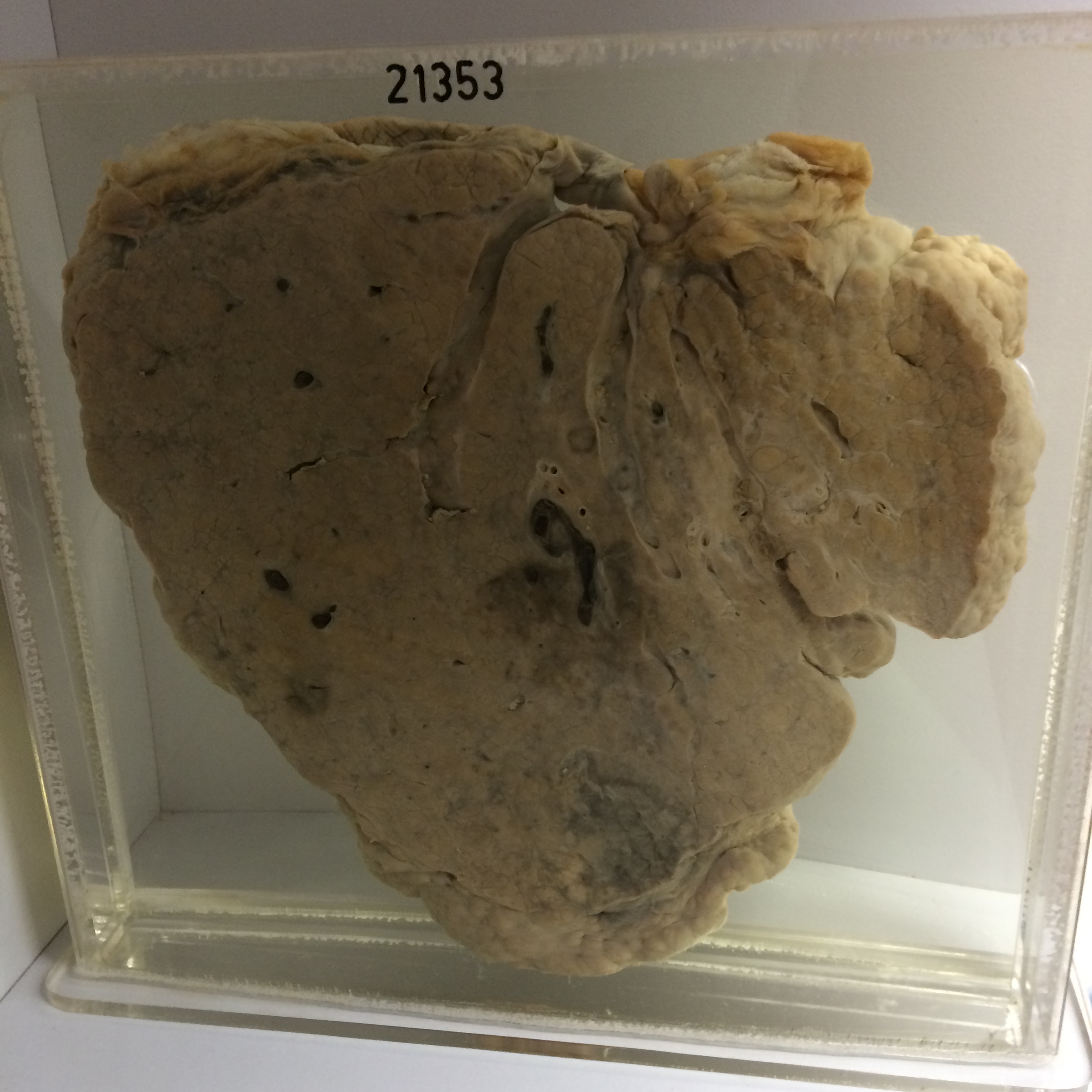
The specimen is the liver. Its surface is markedly and irregularly nodular and the cut surface shows advanced cirrhosis. The regeneration nodules vary in size up to 7 mms in diameter. Dark thrombus-like masses of tumour are present in large branches of the portal vein within the liver. The origin of this tumour in the liver substance is uncertain but may be towards the lower end of the specimen at the inferior border of the right lobe. Histology shows advanced portal cirrhosis with fatty degeneration and diffuse inflammatory infiltration. There is diffuse infiltration by hyperchromatic hepatoma cells with several large bizarre giant cells. Invasion of veins is prominent.