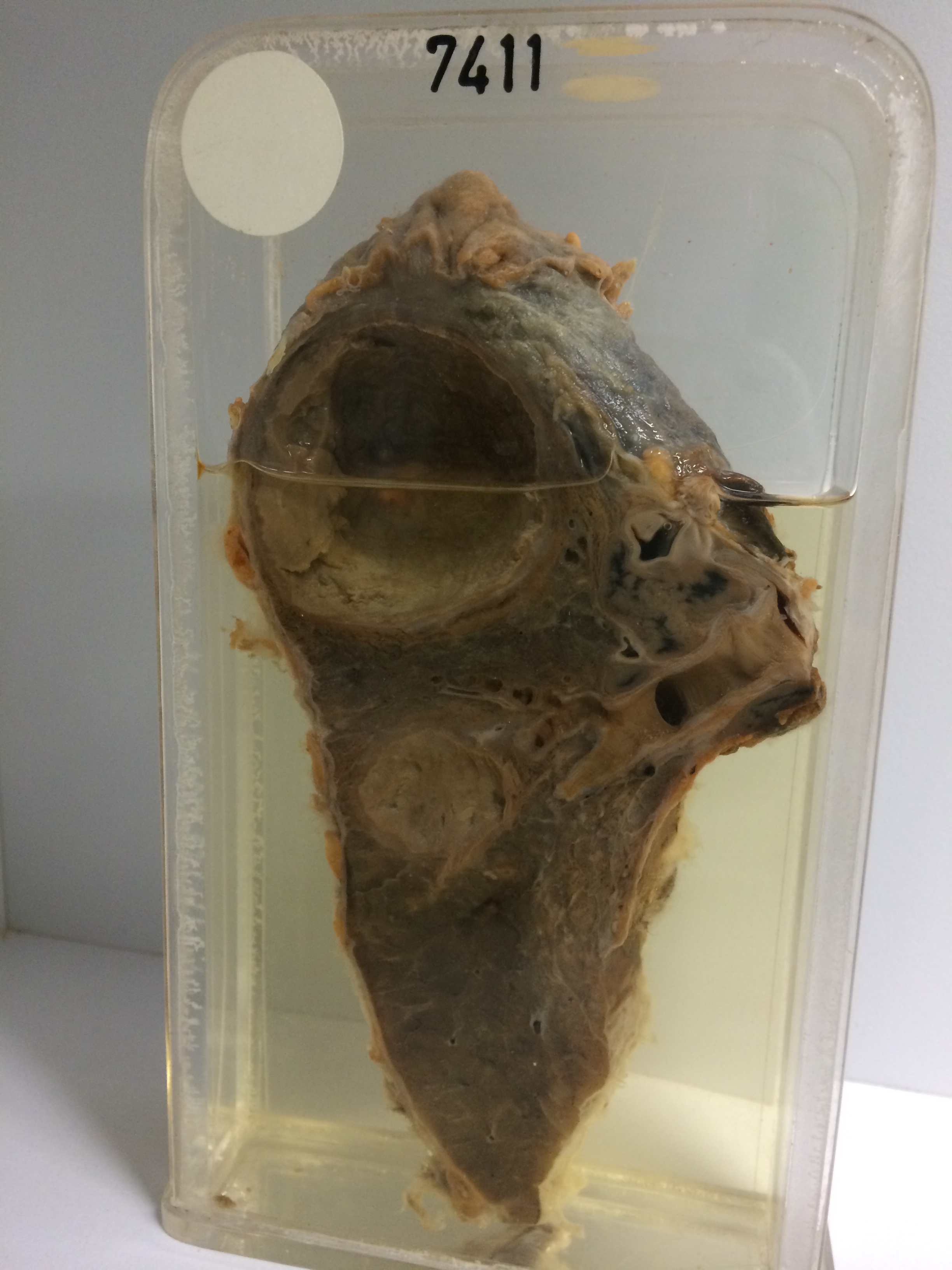
7411 CHRONIC LUNG ABSCESSES
The patient was a man who had right sided pneumonia 17 years previously. There were two recurrences at intervals of about 4 years. One month after the last attack he developed cough with bloodstained sputum and thereafter had several further haemoptyses. X-ray showed an opacity at the right lung base which was cleared by chemotherapy, but a bronchogram showed incomplete filling of the anterior and lateral basal divisions on the right side. These segments were resected.
The specimen shows this resected portion of the lung. At its upper end is a large chronic abscess cavity 5 cms in diameter with thick fibrous walls and containing yellow pultaceous exudate. A rim of lipoid pneumonitis surrounds the capsule of this abscess. A smaller more acute satellite abscess 2.5 cms in diameter is present below the larger cavity. This abscess could be shown to communicate with the bronchus. The reverse of the specimen shows marked fibrotic collapse with bronchiectasis in the lung adjacent to the larger abscess. The overlying pleura is markedly thickened and covered with thin filmy fibrous adhesions. A further area of collapse with fibrosis and bronchiectasis is visible on the medial side of the specimen below the main bronchus.