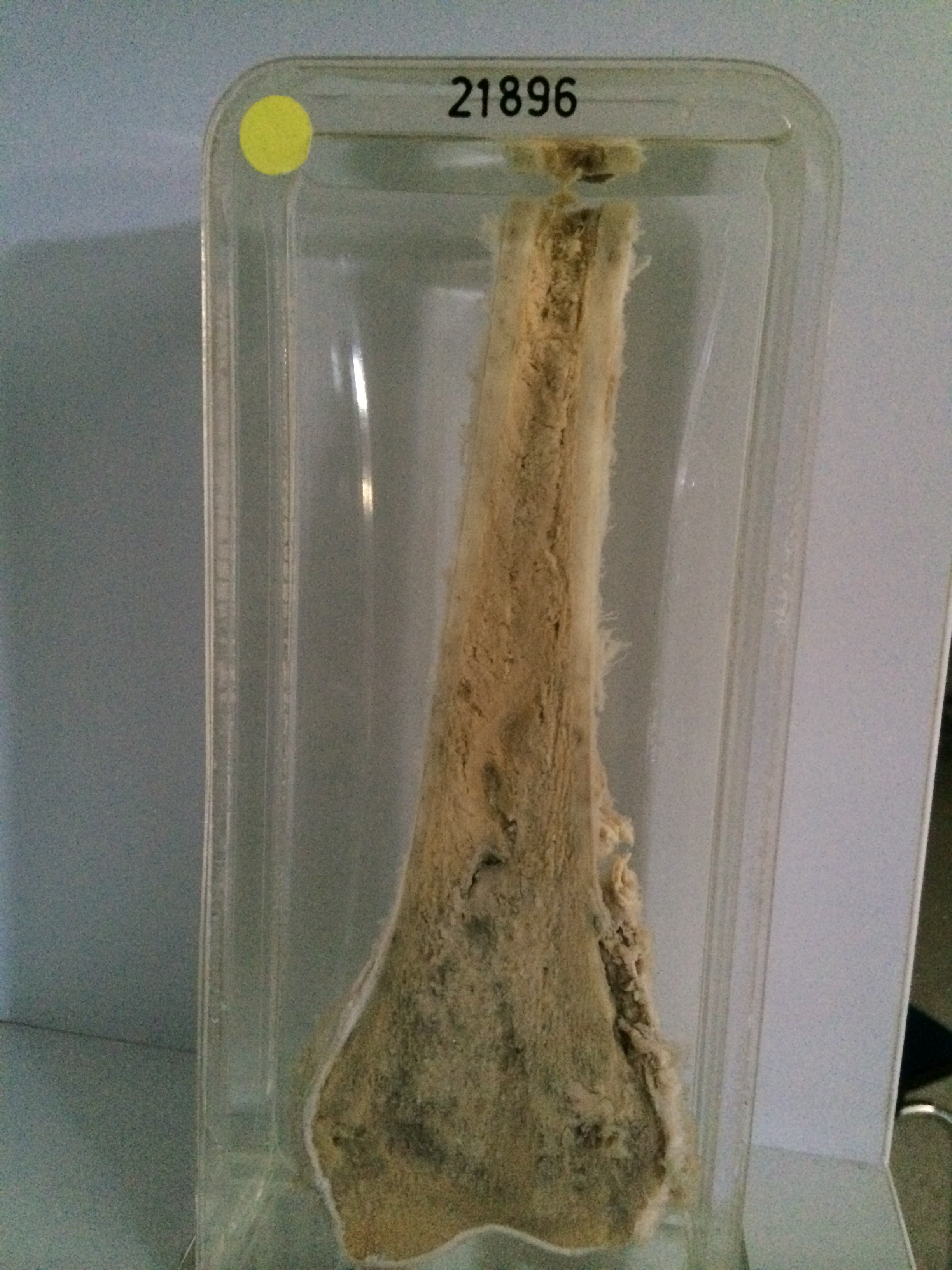
21896 OSTEOGENIC SARCOMA
The patient was a man aged 20 who had a painful swollen knee for 4 months. X-ray showed an osteolytic lesion in the left lateral condyle which was confirmed by biopsy to be an osteogenic sarcoma. The leg was amputated.
The specimen consists of the lower 25 cms of the left femur sectioned in the coronal plane. The marrow at the lower end of the bone shows a patchy haemorrhagic lesion destroying the bony lamella with some extension up to and through the periosteum on the lateral aspect and into the soft tissue posteriorly. Histology showed a typical osteogenic sarcoma with areas of osteoid tissue calcified in places and some areas of spindle cell growth. Mitotic figures were numerous.
Last modified: Monday, 31 July 2017, 11:55 AM