25460 DISSECTING ANEURYSM OF THE AORTA
The patient was a women aged 74 who presented with a history of constant aching chest pain for 4 hours, passing through to the back. The B.P. was 210/105. There was a short harsh systolic murmur at the apex together with a short rough diastolic murmur also at the apex. The right femoral pulse was weak. There was no biochemical or E.C.G. evidence of myocardial infarct and a diagnosis of dissecting aneurysm was therefore made. During the 9 days she survived, the diastolic murmur fluctuated and a harsh systolic murmur continued. At postmortem the body was extensively decomposed, the liver was autolysed and there was bright red staining of the aorta. There was extensive surgical emphysema of the mediastinum, the retropheritoneal mesentery and the anterior chest wall and neck.
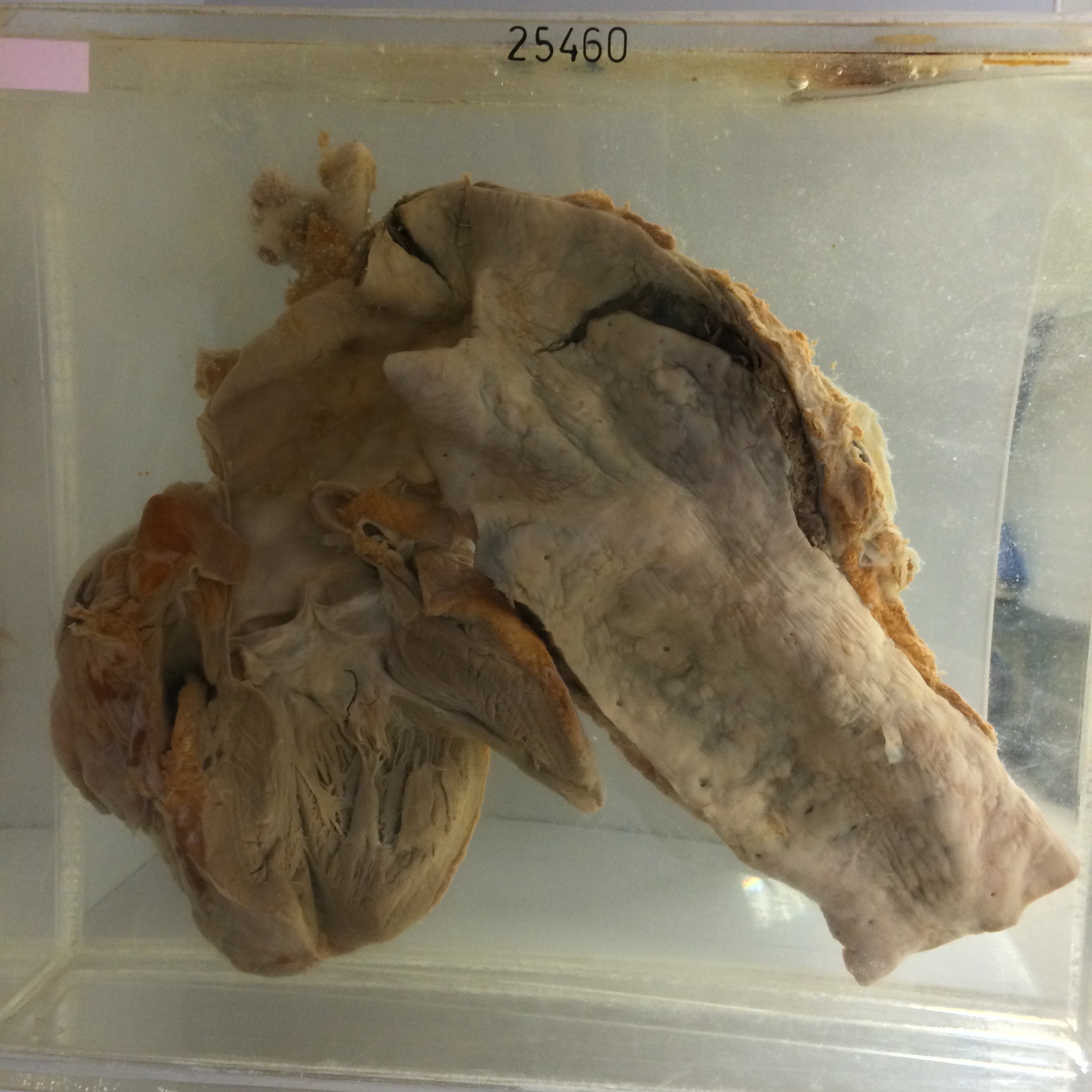
The specimen consists of the lower oesophagus together with the heart. A large superficial ulcer 2.5 cm in diameter is present at the lower end of the oesophagus. It has regular margins and a smooth floor and there is a central perforation measuring 5 x 3 mms which leads into the cavity of the left atrium. The left and right ventricles are both dilated and hypertrophied. There is no record in the history likely that the perforation is artefactual due to postmortem digestion. It seems likely that the ulcer became adherent to the wall of the atrium, and it is possible that during life there was some leakage from it into the mediastinum, but not into the atrium.