21961 FALLOT’S TETRALOGY
The patient was a girl aged 7 who had been cyanosed since birth. She was able to attend school but her exercise tolerance was always reduced. Investigation showed a tetralogy of Fallot with the unusual feature of an anomalous vessel which ran from the right subclavian artery to the right pulmonary artery. There was severe valvular and infundibular obstruction to the outflow tract. She was cyanosed and the fingers were clubbed. There was a right ventricular heave and a loud pansystolic murmur was audible over the anterior chest wall, maximal in the pulmonary area. The B.P. was 120/75. Operation was performed on the bypass with the patient cooled to 30oC. The ventricular septal defect was patched and a large patch was then inserted into the right ventricular outflow and the pulmonary valve area. The patient was returned to the ward in good condition. Next day there was pooling of secretions with increase in cyanosis. She was incubated and the secretions were sucked out. Assisted respiration was continued for six days but respiratory problems persisted and precipitated her death in cardiac arrest on the 7th day. At postmortem there was considerable bronchitis with patches of bronchopneumonia.
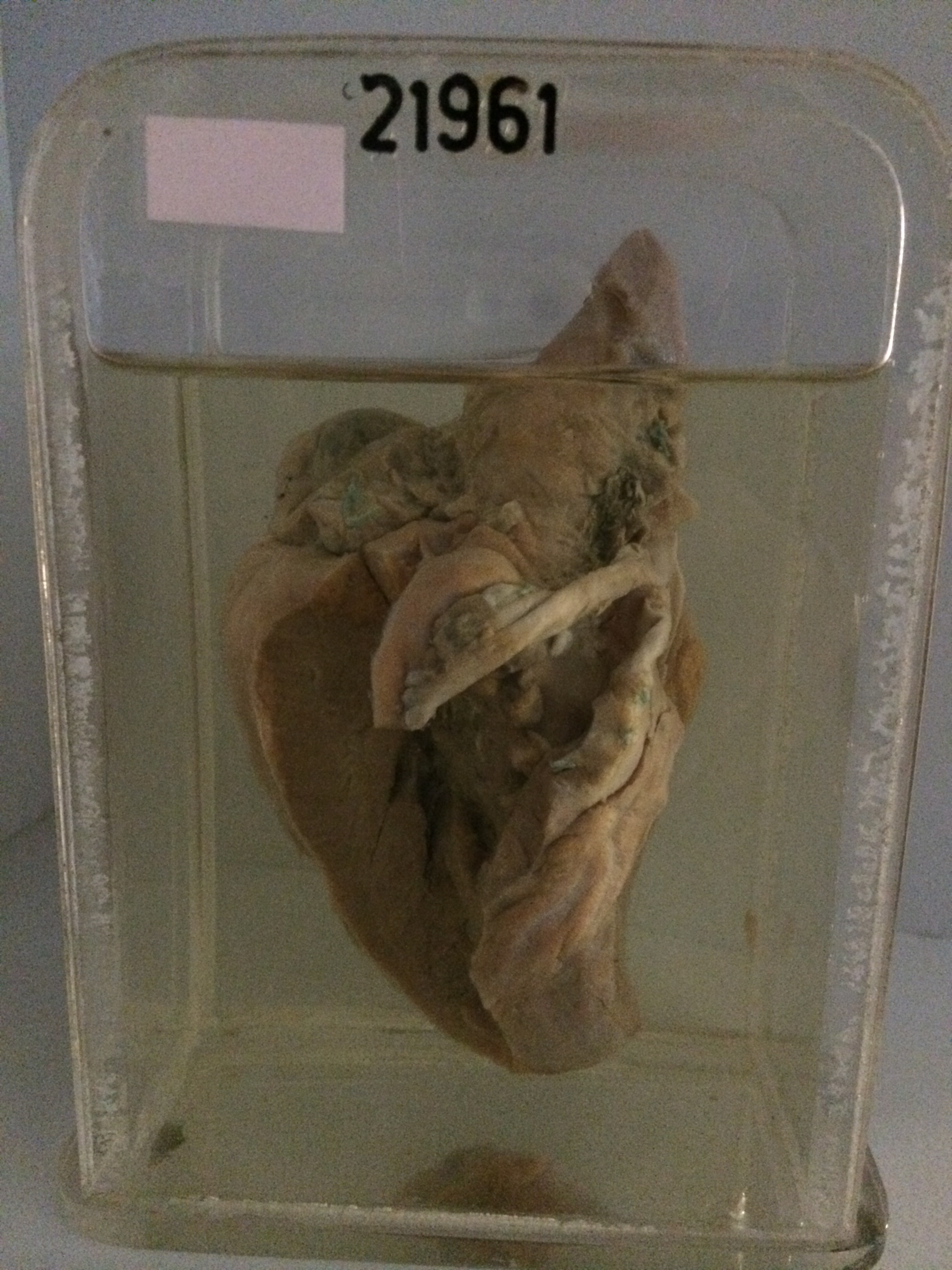
The specimen consists of the heart opened to display some of the above features. The right ventricle is greatly hypertrophied but not obviously dilated. The patch can be seen in place over the ventricular septal defect, with antemortem thrombus and surgical sutures visible on its surface and in the pulmonary outflow tract. The pulmonary valve is absent. The left ventricle is of normal size but is somewhat hypertrophied. The openings of the VSD can be seen above and to the left of the anterior mitral leaflet. The aorta is large and was found to encircle the oesophagus. The left subclavian artery arose from this vessel posteriorly.