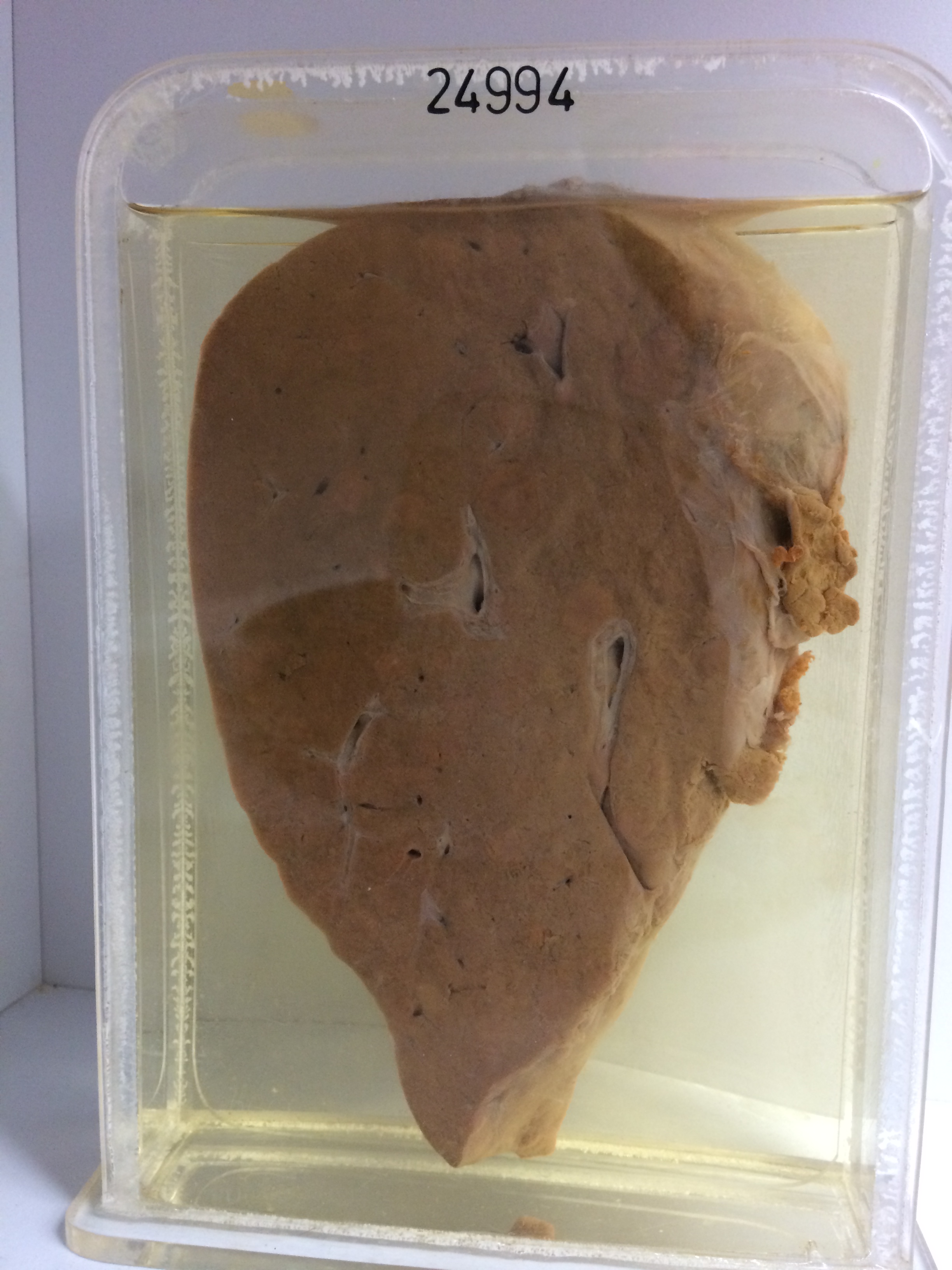
24994 LYMPHOMA
This woman aged 52 presented with vaginal discharge with occasional spotting of blood. The cervical smear was positive for malignant cells. On examination the cervix was normal but there was a hard white ulcerating tumour in the right fornix which rectal examination showed to extend to the right pelvic wall. Biopsy showed lymphosarcoma. Lymphangiograms, chest X-ray and IVP were normal. Megavoltage radiotherapy was given to the pelvis and lower abdomen. Eight months later she returned with a right pleural effusion, ascites and a palpable epigastric mass. She was treated with prednisolone and antimitotic agents but steadily deteriorated and died a few weeks later. At postmortem there was widespread lymphoma involving the peritoneal cavity, bowel, mesentery, liver, spleen, abdominal and mediastinal lymph nodes.
The specimen is a slice of liver which is generally pale. The cut surface is studded with pale round firm lymphomatous infiltrates varying in size up to about 1.5 cms in diameter. The nodules are mainly discrete but are becoming confluent in a few places. Superficial nodules project as smooth rounded swellings on the surface of the liver. The intervening liver substance shows some zonal congestion. Histology shows a pleomorphic reticulum-cell sarcoma poor in lymphocytes and with many multinuclear giant cells.