23380 SMALL PRIMARY CARCINOMA
The patient, an 80 year old man, presented for investigation of a ‘coin’ lesion noted on a routine chest X-ray. He complained of some weight loss over the previous six months and progressive weakness, involving especially the legs and suggesting a proximal muscular disorder. He had smoked 15 to 20 cigarettes for many years and had a rather persistent cough which had been more pronounced for three years. He denied any recent change in cough or sputum production. There was no dyspnoea, haemoptysis or chest pain. He died suddenly before the nature of the lesion could be investigated.
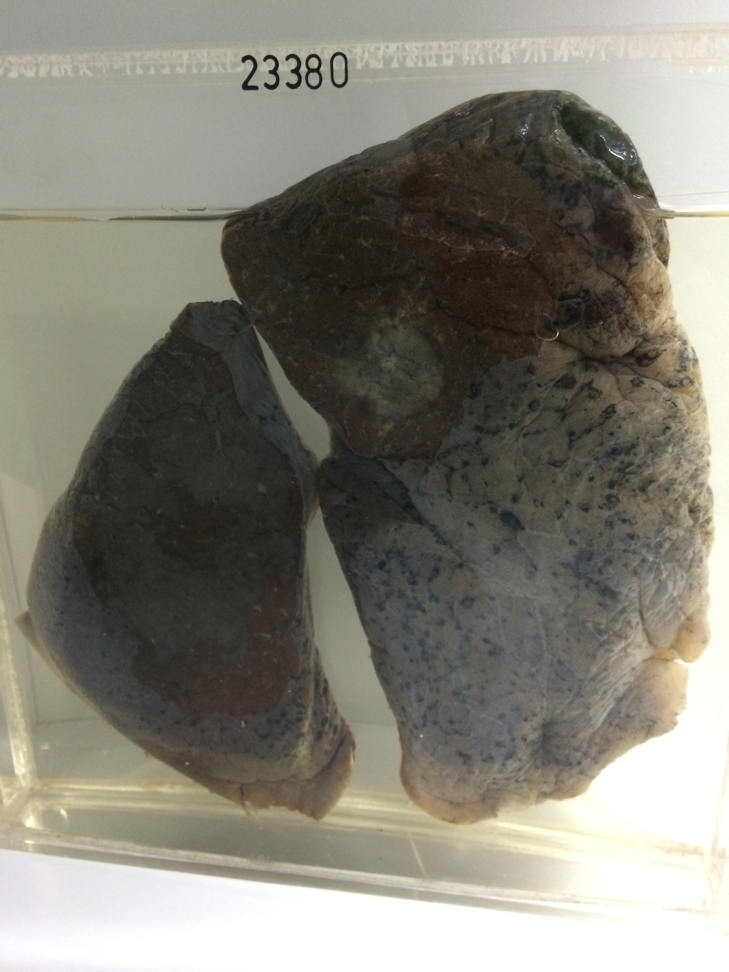
The specimen, consisting of part of the right lung, shows a rounded neoplasm 3 cms in diameter in the upper lobe. It has an irregular infiltrated edge which is penetrating the surrounding congested lung. There is generalised mild chronic venous congestion and some acute terminal congestion in the lower lobe. At this age almost all ‘coin’ lesions noted on a chest X-ray in an asymptomatic patient are malignant. Histology shows a moderately well differentiated squamous carcinoma.