23790 SQUAMOUS CELL CARCINOMA AND RADIATION NECROSIS
The patient, a 60-year old man, had a squamous cell carcinoma on the dorsum of the left hand for 11 years. He had various unsuccessful treatments including radiotherapy and local surgical removal but eventually amputation was required.
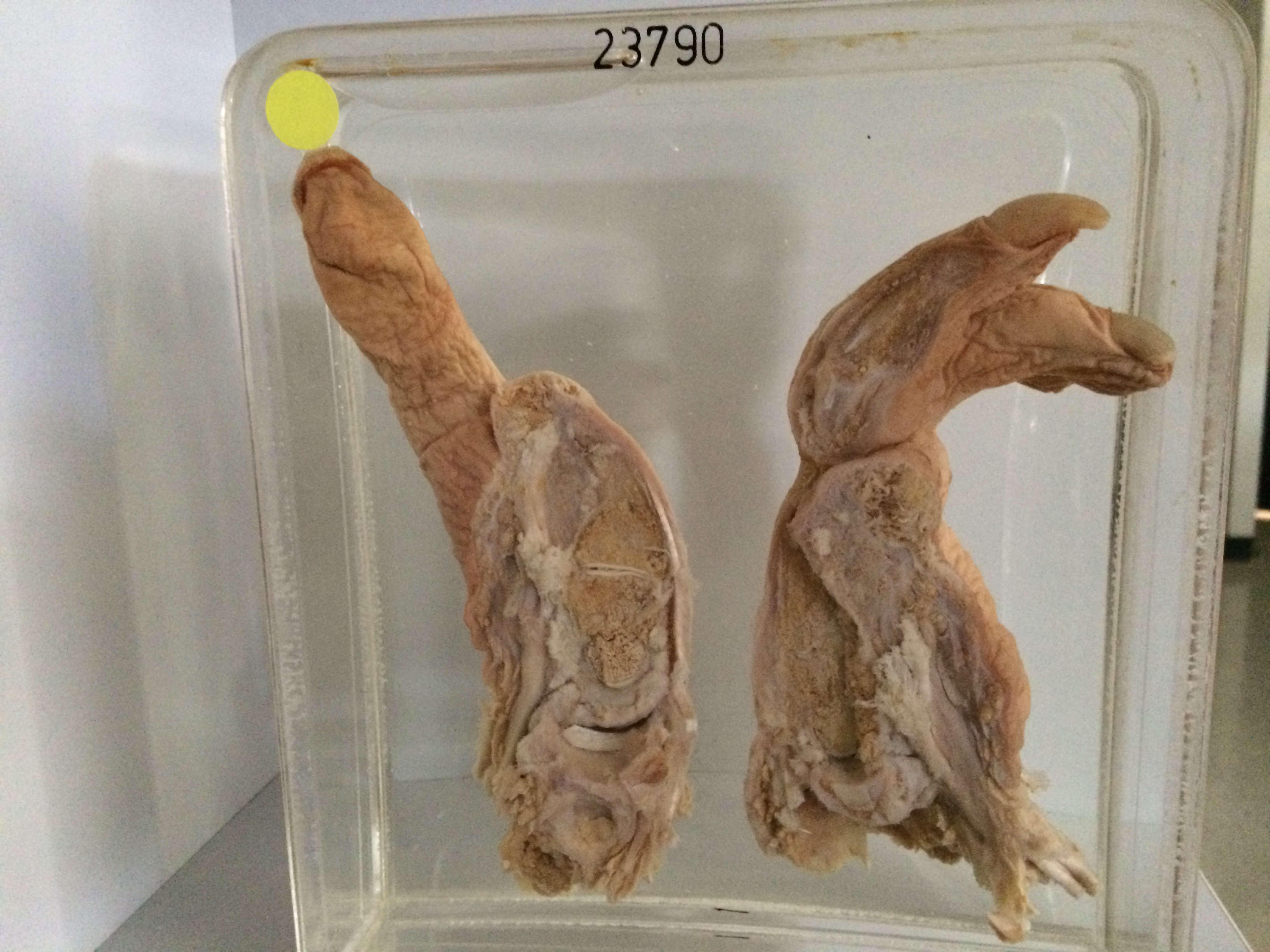
The specimen consists of the left hand bisected between the thumb and forefinger (the index finger has been amputated). On the dorsum of the hand close to the line of incision there is a hyperkeratotic elevated nodule 1.5 cms in diameter. There is a similar lesion in the region of the anatomical snuff box. The skin of the dorsum of the hand is scaly and atrophic and there are the orifices of two sinuses. At the base of the ring finger there id a further hyperkeratotic lesion. The skin of the fingers is also scaly and atrophic. The section reveals necrotic and disorganised metacarpal bones. Histology of the hyperkeratotic nodules showed squamous cell carcinoma. Histology of the dorsum of the hand showed epidermal atrophy, chronic inflammatory cell infiltration and oedema and fibrosis of the subcutaneous fat. The small vessels were narrowed and their walls thickened and hyalinised. These findings are consistent with radiation necrosis.