17991 CRYPTOCOCCOSIS (TORULOSIS)
The patient was a male wood merchant aged 64. Three months previously a medical examination by his local doctor revealed no abnormality. A month later he developed constant bifrontal headache. Two weeks later two facial nodules developed which resembled basal cell carcinoma but grew more rapidly. One of these nodules ulcerated and was excised. Histology revealed torular infection. He was accordingly admitted to hospital and the CSF was found to contain torula although there were no localising neurological signs. Chest X-ray showed a rounded opacity in the right lung. He was treated by Amphotericin B and on the 11th day the remaining facial lesion and the right lower lung lobe were excised. He made an uneventful recovery.
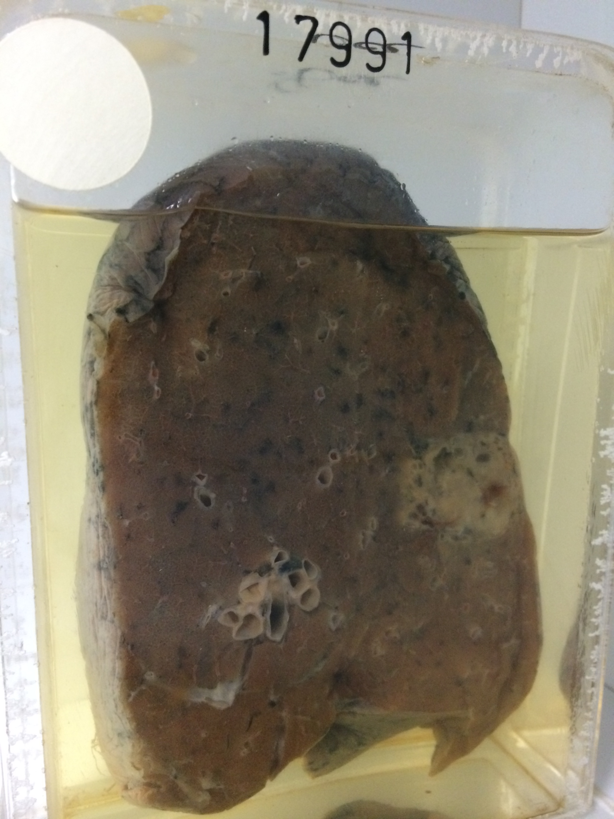
The specimen consists of the right lower lobe sectioned to show a circumscribed rounded mass 3 cms in diameter lying just beneath the pleura. The cut surface is grey with some gelatinous areas and some small areas of haemorrhage and cystic degeneration. The border is rather wavy and the mass has no definite capsule. There is some lipoid pneumonitis in the lung immediately medial to the mass. The remainder of the lung is normal. Histology shows typical torula with a granulomatous inflammatory response.