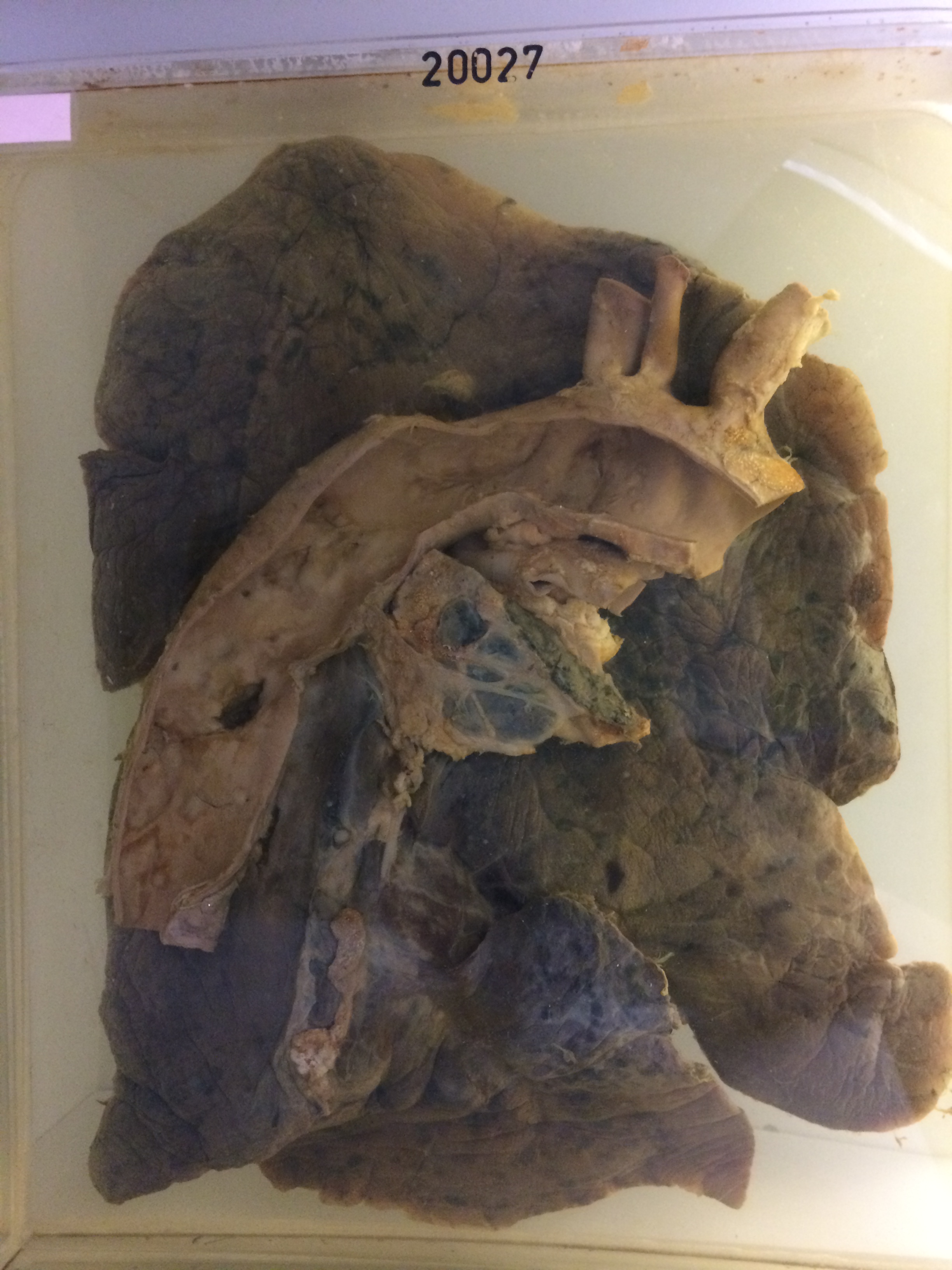
20027 PULMONARY TUBERCULOSIS WITH EROSION OF AORTA
This man aged 77 had a dry cough for 4 months. Ten days before admission there was a small haemoptysis and on the day of admission there were two haemoptysis each of some 200 mls. A further haemoptysis occurred two days later and he died on the 4th day after a further very large haemoptysis. Chest X-ray while in hospital showed a mass at the level of the left hilum posteriorly.
The specimen consists of the left lung and portion of the thoracic aorta. There is a pale tuberculous mass some 4 cms in diameter in the apical segment of the left lower lobe. Just beneath this there is a wedge-shaped area of more acute tuberculosis extending out to the pleura posteriorly. Yellow caseous exudate is present in the centre of the mass. The medial aspect of the lesion is adherent to the descending thoracic aorta and an irregular split 1½ cms in length can be seen penetrating the full thickness of the vessel. Hilar glands are involved by old tuberculosis disease. Histology shows active fibro-caseous tuberculosis with a good deal of granulation tissue and some areas of caseation.