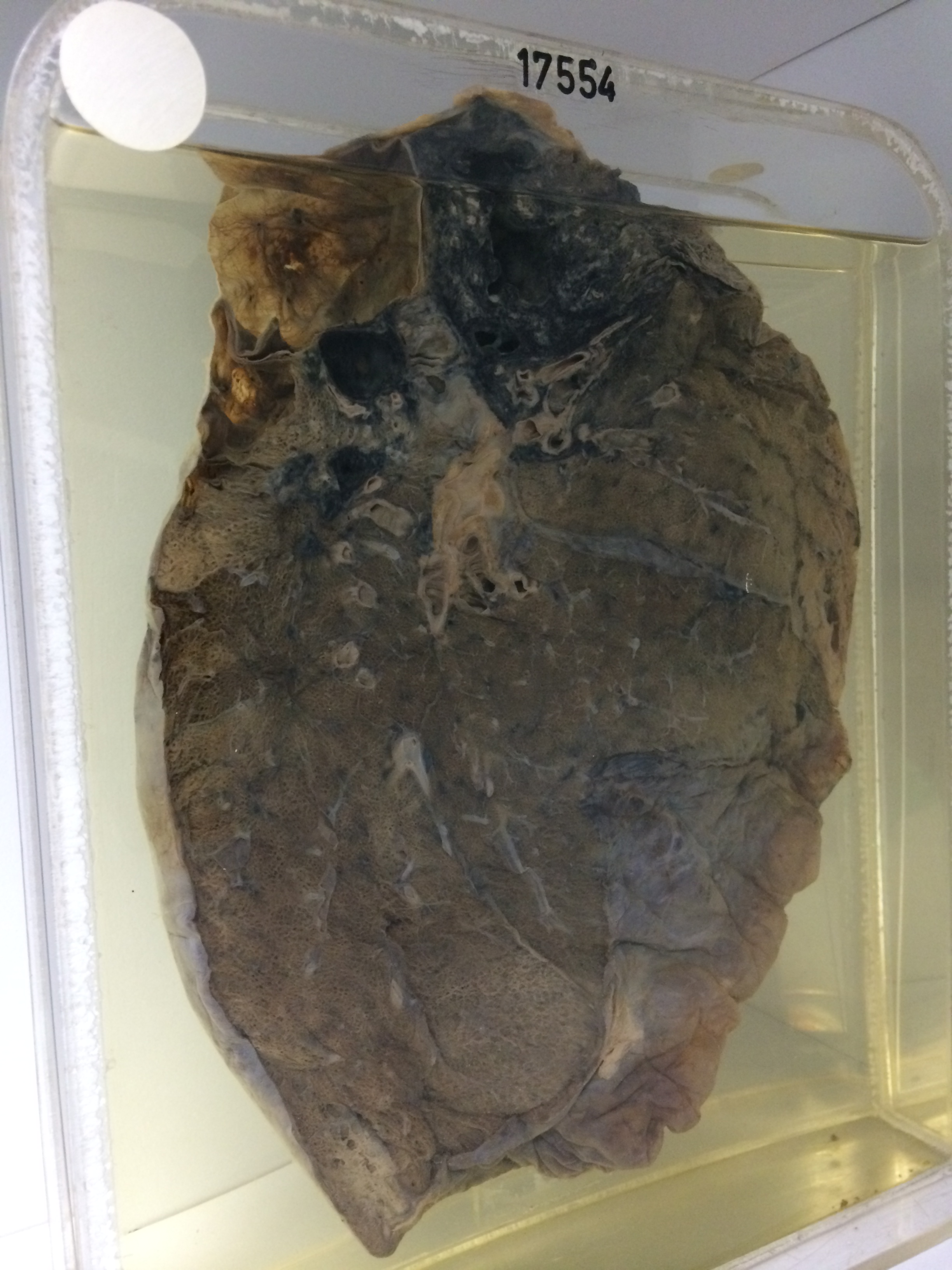
17554 SILICOSIS WITH CAVITATION
The patient was a man aged 86. He had a myocardial infarct 2 years previously. On this occasion there was dyspnoea and a chronic productive cough. Sputum culture grew a pneumococcus. There was a leucocytosis of 28000 neutrophils/cu.mm. He was treated with penicillin and improved initially, but died on the 4th day. Unfortunately there is no record of his occupational history. At postmortem the aortic valve was calcified but the right ventricle was not obviously hypertrophied.
The specimen consists of the left lung sectioned to show a large apical silicotic mass densely pigmented by included carbon and showing two ragged central cavities, the largest of which measures 2 cms in diameter. These cavities have relatively smooth walls. The overlying pleura is thickened and fibrous. There is a very large emphysematous bulla 5 cms in diameter in the posterior subapical region just behind this fibrotic mass. A smaller silicotic nodule 1 cm in diameter lies below this emphysematous region. The remainder of the lung appears normal. In particular there is no focal emphysema or focal anthracosis. There is a zone of septic pneumonic consolidation along the anterior border of the upper lobe with some overlying pleurisy. Histology shows typical silicosis. There is no evidence of tuberculosis.