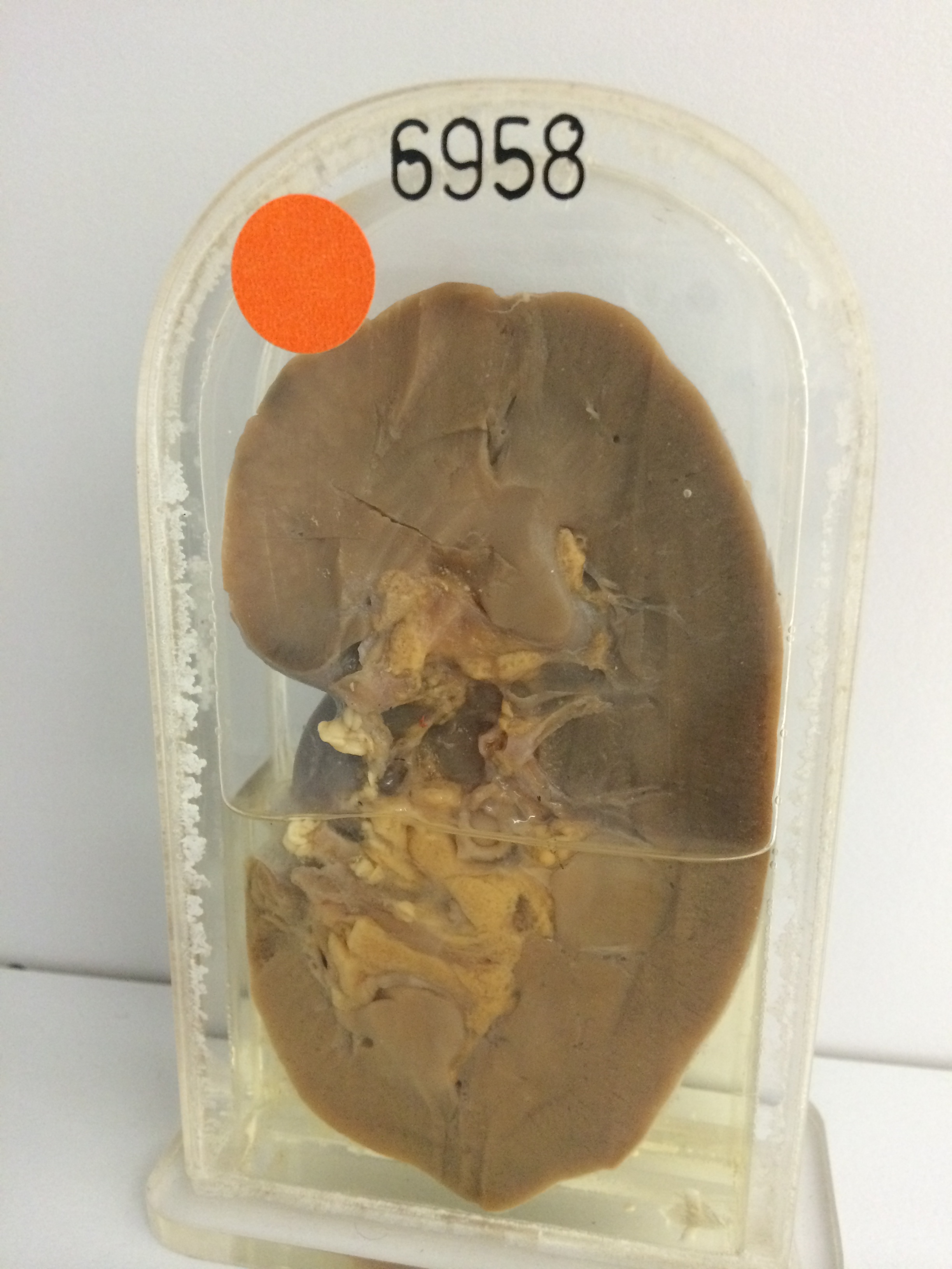
6958 RENAL TUBERCULOSIS
The patient was a woman who developed urinary frequency, dysuria, haematuria and pain in the right renal angle shortly after an operation for the repair of a cystocoele. Cystoscopy showed chronic diffuse cystitis and the right ureteric orifice was incompletely closed, presenting a partial ‘golf-hole’ appearance. Intravenous pyelogram showed slight distortion in the upper calyces of the right kidney. Pus cells and many tubercle bacilli were found in the urine. She was treated with streptomycin for a month. The kidney was removed 6 months after the first operation because there was still loin pain and dysuria though urinary cultures were negative.
The specimen shows a coronal section of the kidney which measures 11 cms in length with a smooth surface and a normal-appearing cortex and medulla except at the upper pole. At the upper pole there is diffuse tuberculous disease in the cortex where many conglomerate tubercles are present. The medullary pyramid in this region is not quite in the plane of section but seems only slightly affected. Histology showed typical tubercles in varying stages of maturity including many with caseating centres and giant cells.