6882 VIRAL HEPATITIS WITH MULTIPLE NODULAR HYPERPLASIA
The patient was a woman aged 22 whose illness lasted 3 years. It began with a generalised skin rash accompanied by slight jaundice, pale motions and dark urine. These symptoms and signs continued for two years and then disappeared completely for 9 months. For 5 months before her last admission there was anorexia with increasing jaundice. The urine was deeply pigmented but the faeces appeared normal. A moderate haematemesis occurred 4 months before admission and there was ascites for the last week. Paracentesis yielded clear bile stained fluid. The prothrombin level was 20%, serum alkaline phosphatase was normal, cephalin cholesterol markedly positive. Repeated haematemeses with melaena occurred and she died on the 10th day. At postmortem oesophageal varices were present and there was blood in the bowel. The liver was small and soft. The spleen weighed 670 gms and was firm, fibrous and congested.
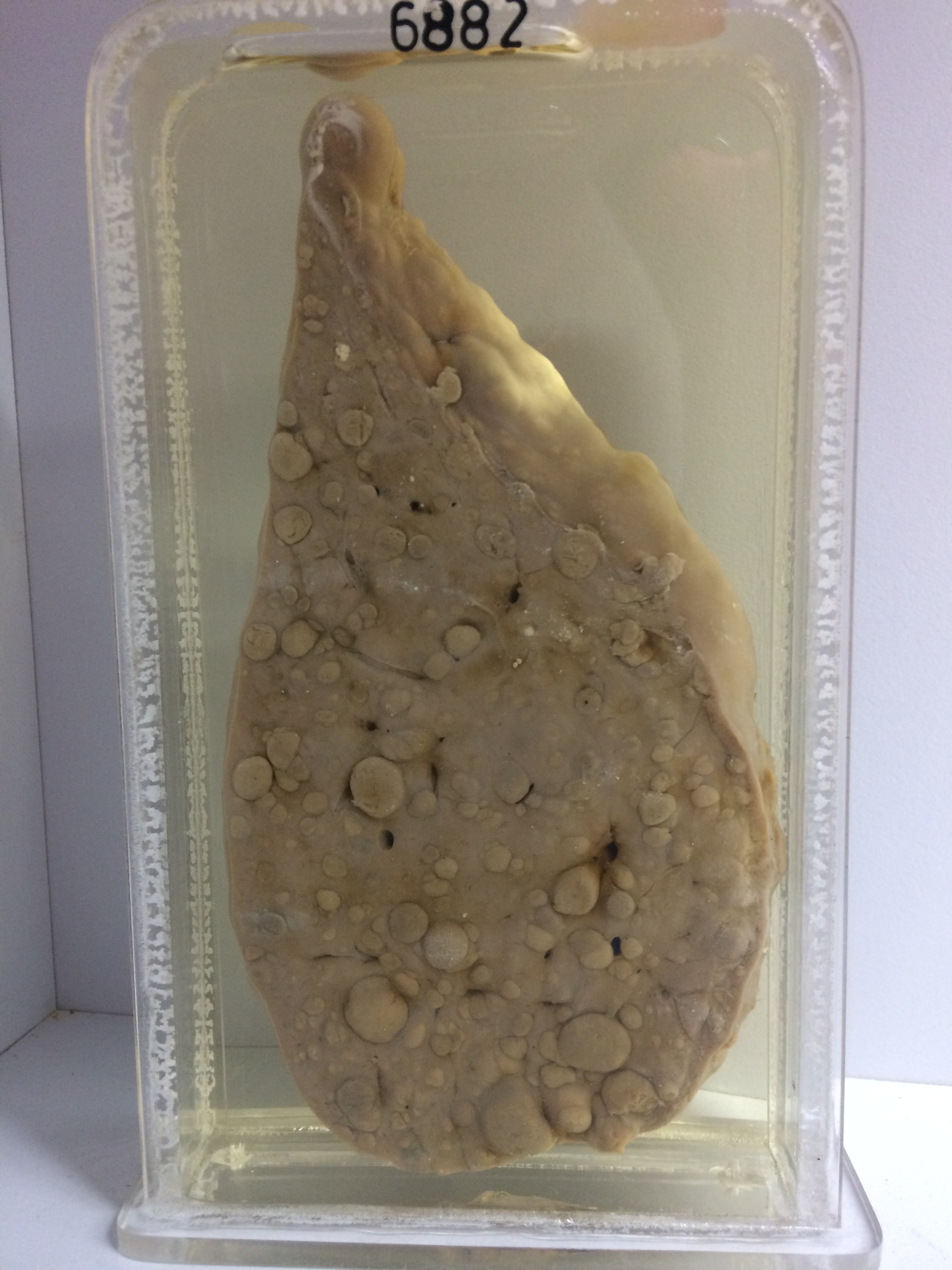
The specimen is a slice of liver. Gross nodules up to 2 cms in diameter protrude on the surface. The cut surface shows large bile stained regeneration nodules of liver tissue up to 1.5 cm in diameter. Large areas of hyaline fibrous tissue intervene between the nodules. Histology shows large irregular islands of hypertrophied liver cells, most of them showing intense cloudy swelling. There is some bile retention in some places and there is moderate proliferation of small bile ducts in the zone of intervening fibrosis. Inflammatory cells are relatively sparse.