16703 PULMONARY INFARCTS WITH CAVITATION
The patient was a man aged 69 with a 12-months history of breathlessness. The ankles had been swollen for 2 months and there had been cough for 2 weeks accompanied by pain in the right side of the chest and bloodstained sputum. On examination the JVP was raised. There was a lumbar pad and the legs were swollen to the knees.
The B.P. was 95/85 and there was a loud systolic murmur maximal at the left sternal edge and radiating to the neck. X-ray confirmed calcific aortic stenosis and also showed an opacity in the lower right lung field. Pulmonary infarction was diagnosed. A few days later he became jaundiced with a bilirubin of 6 mg%. Urinary output fell and he gradually deteriorated until his death on the 12th hospital day. At postmortem there was marked calcific aortic stenosis with left ventricular hypertrophy. The right atrium was markedly dilated and the right ventricle was moderately dilated and hypertrophied. Antemortem thrombus was present in the left atrial appendix and in both femoral veins. There was a large bloodstained right pleural effusion.
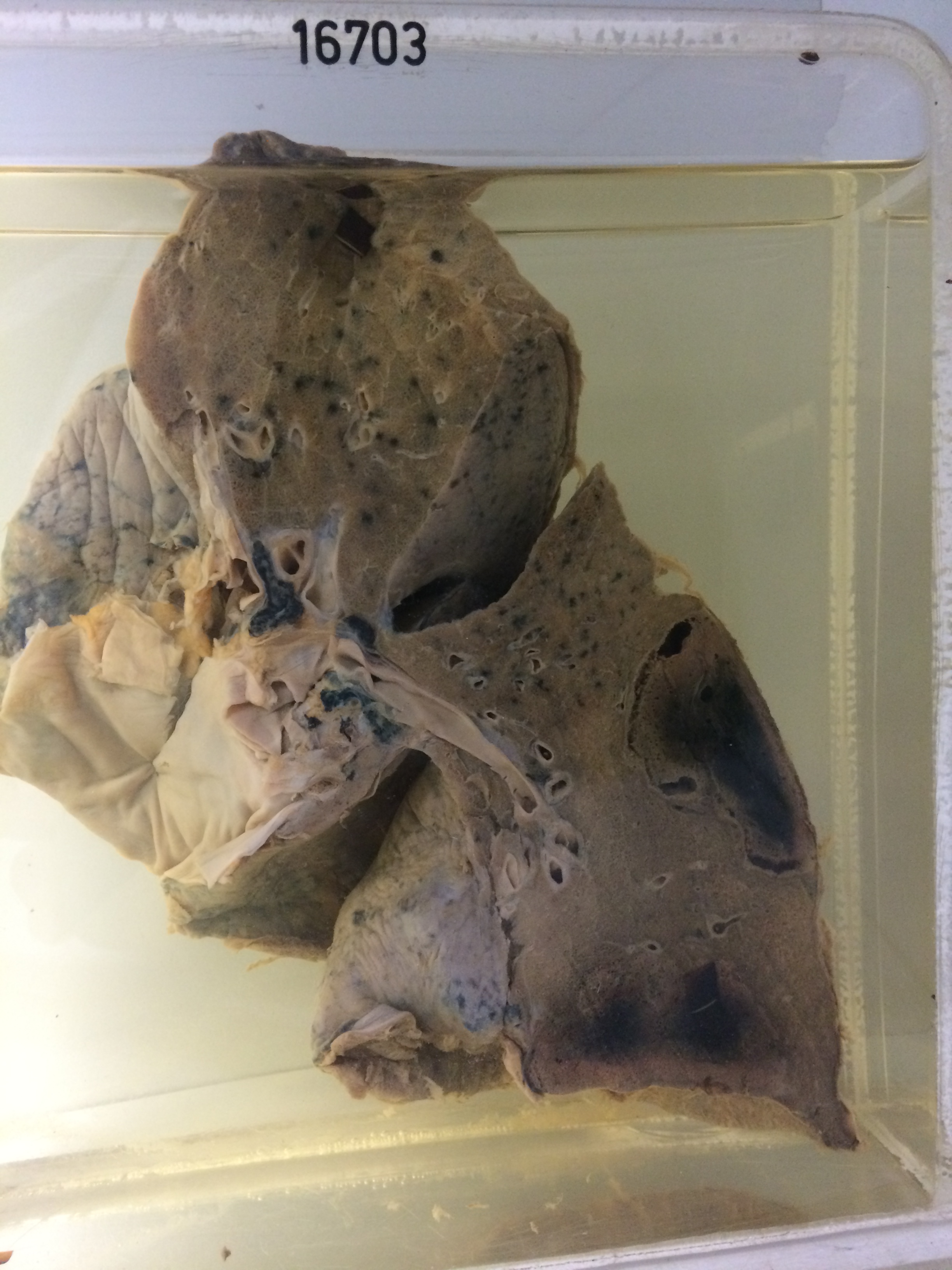
The specimen consists of right lung sectioned to show two massive haemorrhagic infarcts in the lower lobe. The infarcts are wedge-shaped with their bases on the pleura. They are of some duration and autolytic softening has progressed to the stage of cavitation which is most marked in the upper infarct. There is overlying fibrinous pleurisy. There was marked chronic passive venous congestion of liver and kidneys. It was concluded that the jaundice was at least partly due to autolytic destruction of red cells in the pulmonary infarcts.