15793 LOBAR PNEUMONIA
A man aged 46 had been unwell for 4-5 weeks with cough which became worse 3 days before his admission. There was some pleuritic pain and slight haemoptysis on the day of admission. He was then cyanosed and febril with rapid respiration and pulse. There were clinical and X-ray signs of bilateral upper-zone pneumonia, worse on the right. Treatment with penicillin was begun. Sputum culture did not show a pathogenic organism. The fever rapidly resolved but the patient continued to deteriorate with persistent low blood pressure and he died 24 hours after admission. At postmortem the only significant findings were in the lungs and in the heart where there was pallor of the myocardium with moderate dilatation of the right ventricle. Gram-positive cocci were seen in smears from the lungs but did not grow on culture.
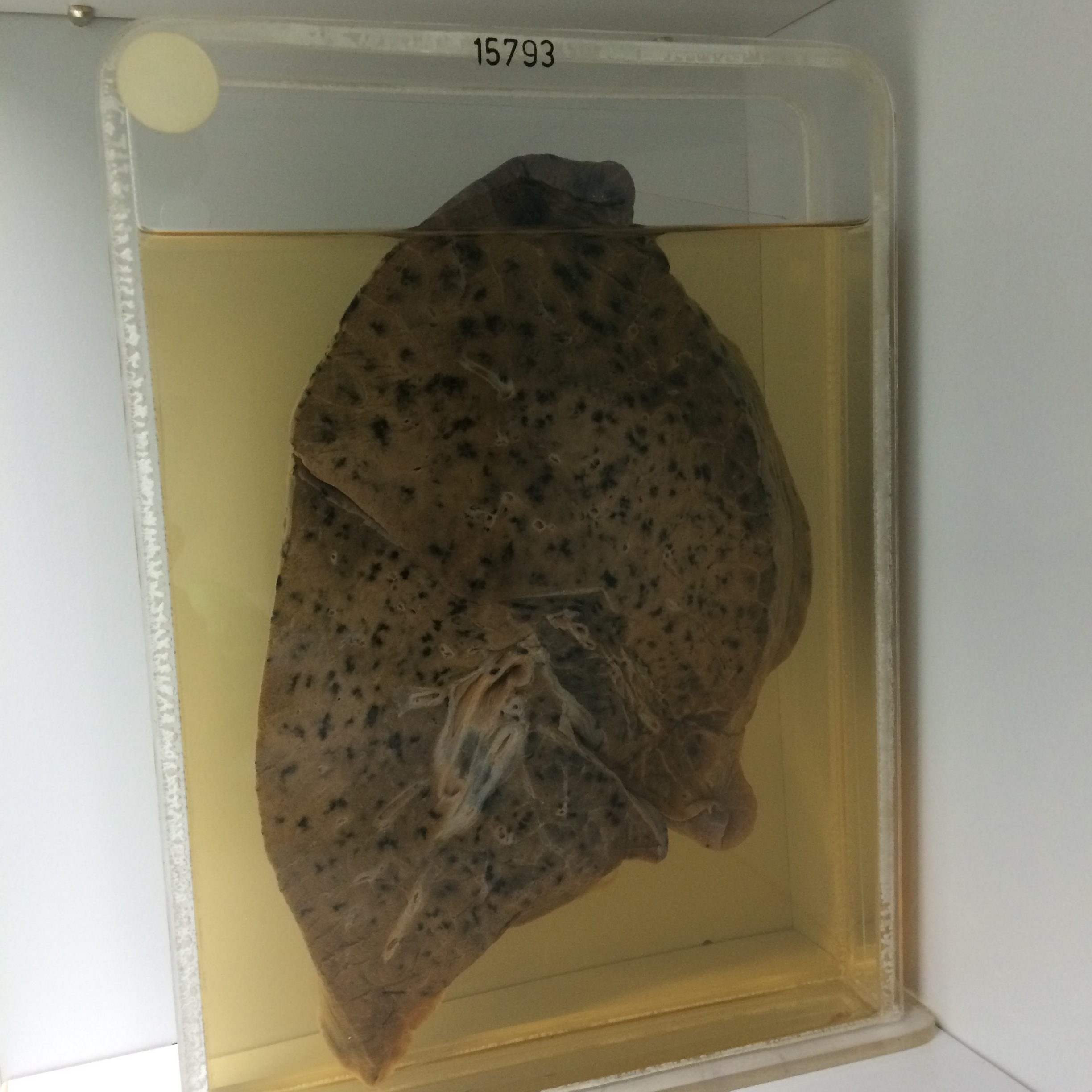
The specimen consists of a slice of the right lung showing ill-defined grey hepatization affecting the posterior and lingular segments of the upper lobe and most of the lower lobe. There is also moderate focal centrilobular emphysema with anthracosis. The overlying pleura shows very little reaction. Histology shows lobar consolidation with a mixed polymorph and mononuclear exudate. Alveolar walls are congested and there are many small areas of haemorrhage.