17262 BACTERIAL ENDOCARDITIS OF MITRAL VALVE
The patient was a man aged 50. Eighteen days before admission he developed a sudden severe pain in the left hip. The pain moved down the leg to the foot which became hot, swollen and tender. Two weeks later a sudden pain occurred in the shoulder and arms. On examination the B.P. was 160/80, the temperature was 39oC and he was cyanotic. A loud systolic murmur was present at the apex and was transmitted to the axilla. The liver was enlarged 3 fingers below the right costal margin. The provisional diagnosis was of subacute bacterial endocarditis viridans. Treatment with intramuscular penicillin was begun. Later he developed abdominal distension with pain and vomiting, diarrhoea and melaena.
The serum amylase was considerably raised and he was treated as acute pancreatitis with drip and suction. Meanwhile he became jaundiced and the diarrhoea worsened while the abdomen remained distended. He was managed conservatively until the 14th day after admission when laparotomy showed mesenteric infarction and massive gangrene of the small gut with multiple perforations. Almost all the small gut was resected and a duodenoileal anastomosis was performed. After the operation the right foot became cyanosed and on the 4th day the abdominal wound broke down. He continued to deteriorate and died on the 14th day after the operation. At postmortem there was gangrene of the toes of the right foot, bilateral large pleural effusions, general peritonitis, embolic occlusion of the superior mesenteric artery and bilateral subphrenic abscesses.
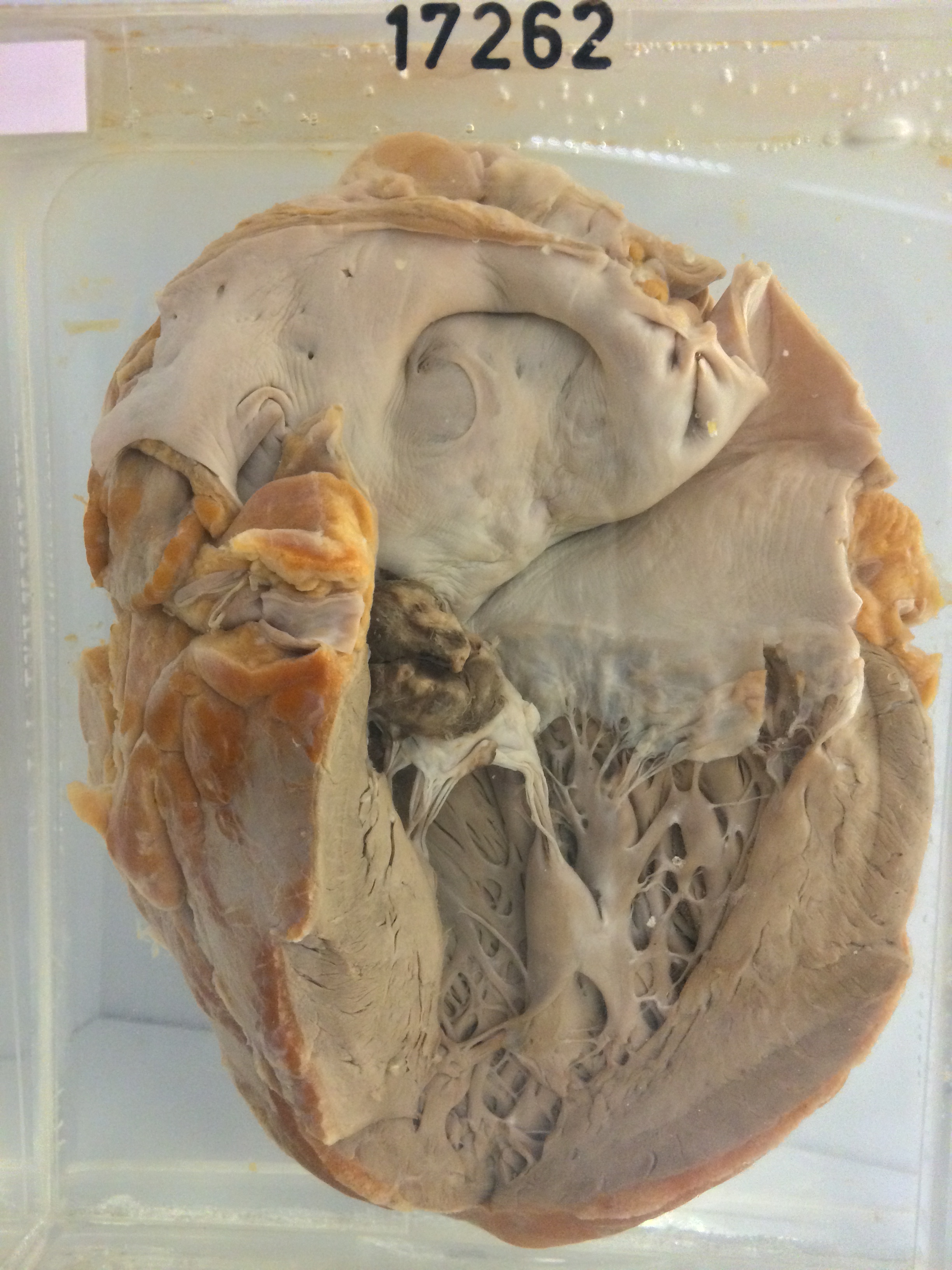
The specimen consists of the heart opened to display the left side. A large, rather smooth and dome-shaped vegetation is present on the anterior leaflet of the mitral valve. The vegetation measures about 2.5 cm x 2cms and is about 1 cm high. A probe could be passed at postmortem through a small perforation of the valve cusp at the base of the vegetation. A further small vegetation is present at the left end of the posterior mitral cusp is thin and shows no evidence of previous rheumatism. The anterior cusp is thicker and appears somewhat scarred, possibly indicative of a rheumatic origin.