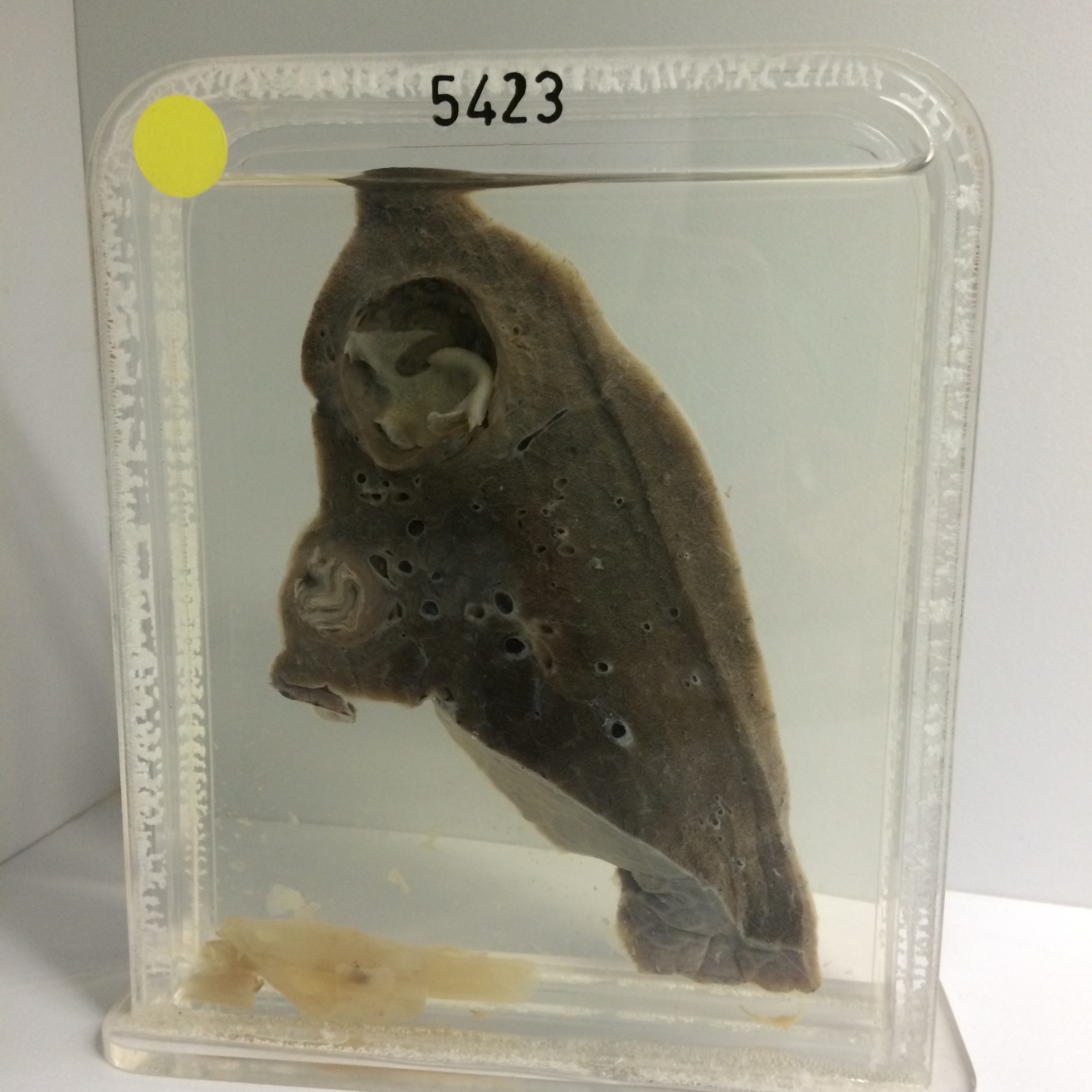
5423 HYDATID CYSTS
The only information available is that the patient was a woman who had an irritating cough with repeated haemoptysis. X-ray shows two rounded opacities in the right lower lobe and another in the left lung. There was an eosinophilia of 7%. The right lower lobe was resected.
The specimen shows the right lower lobe cut to display the two hydatid cysts. One measures 3 cms in diameter and the other 1 cm which has a relatively thin fibrous capsule and contains collapsed laminated membrane. The remainder of the lobe appears normal apart from some antemortem thrombi in medium-size vessels, perhaps related to the surgery.
Last modified: Thursday, 3 August 2017, 9:40 AM