10823 BILATERAL HYDRONEPHROSIS
The patient was a woman aged 47 who had both ureters transplanted into the sigmoid colon at the age of 16 for incontinence. The operation was performed by Sir Henry Newland. Ten months before her death portion of the sigmoid colon was resected for carcinoma and end-to-end anastomosis was performed. At this operation the left kidney was thought to be non-functional and the left ureter was tied. Thereafter she had recurrent severe lower abdominal pain with vomiting. It was thought that chronic intestinal obstruction might have been the cause of the renal insufficiency and laparotomy was performed. Multiple adhesions were divided and several feet of small bowel were excised. However her condition gradually deteriorated and just before death the BUN was 280 mg%. At postmortem mucinous metastases of sigmoid carcinoma were found in the serosal coat of the bowel and in the mesentery.
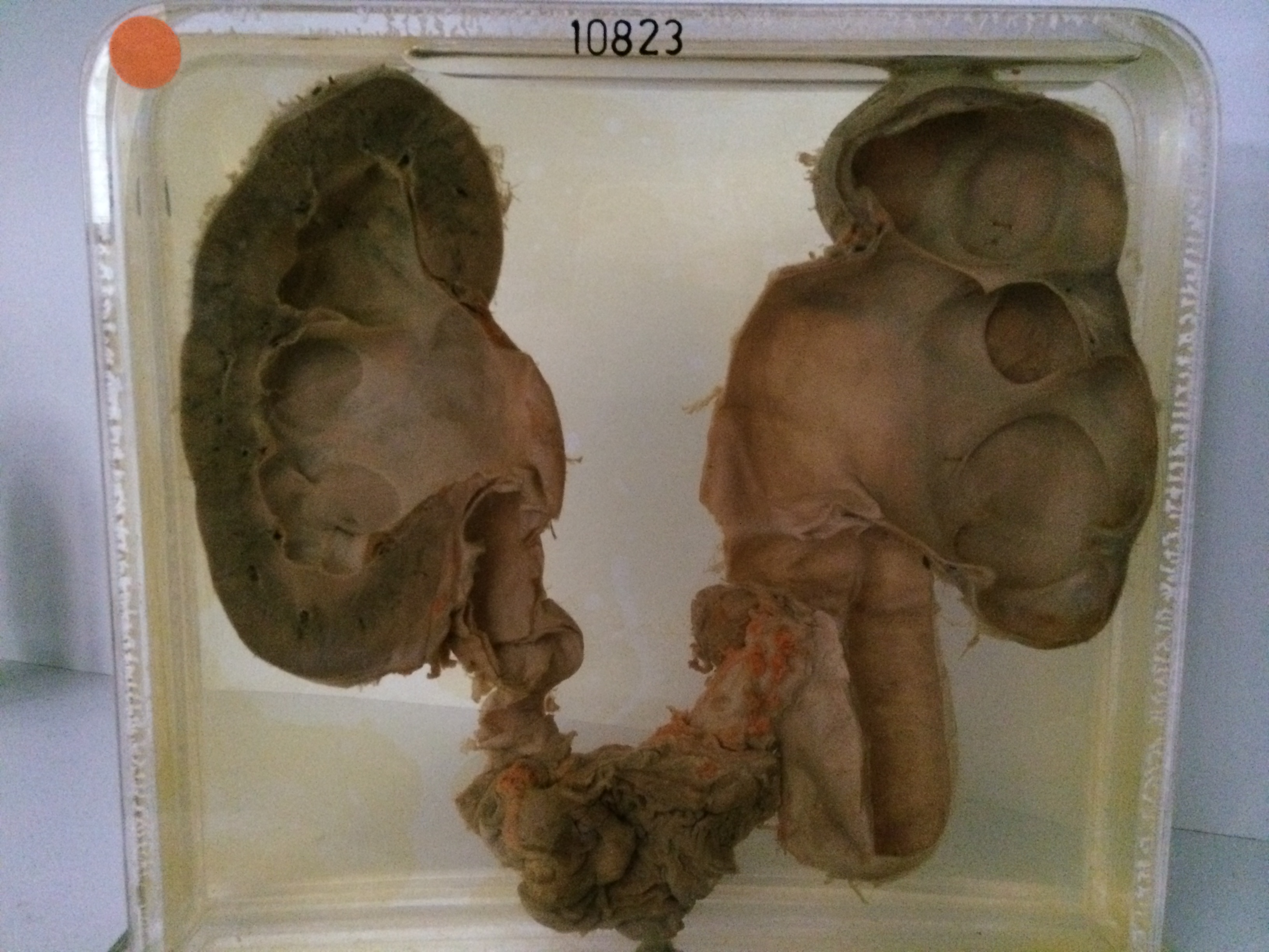
The specimen consists of coronal sections of both kidneys. The left kidney shows gross intrarenal and extrarenal hydronephrosis and hydrocalycosis. Only a thin rim of renal tissue remains. The ureter is also grossly dilated, fibrous and tortuous and has a diameter of 2 cms. The right kidney measures 13 cms in length and shows marked intrarenal and extrarenal hydronephrosis but a good mass of renal tissue survives. The right ureter has a diameter of 1.5 cms and is dilated and tortuous. The colonic insertion of the ureters is shown at the bottom of the specimen. Recurrent tumour is visible surrounding the entrance of the right ureter producing a nodule that projects into the intestinal lumen. Histology of the right kidney shows mild diffuse interstitial fibrosis, but there are very few hyaline glomeruli. The surviving glomeruli are large and slightly hypercellular, but show little abnormality. There is little evidence of significant pyelonephritis.