25441 ALCOHOLIC CARDIOMYOPATHY
The patient was a man aged 51 who had been suffering from heart disease for 6 years. He formerly drank 1 bottle of wine per day and a great deal of beer, but had been drinking less in the last 6 years. There was a 3 year history of gout controlled by treatment. At his last admission there was epigastric pain and nausea, sometimes associated with vomiting. There was marked guarding in the epigastrium and the liver was enlarged and tender. The cardiac apex was in the 5th space lateral to the midclavicular line. Chest X-ray showed pulmonary oedema, and there was ECG evidence of an old antero-septal infarct. The epigastric pain suddenly became more severe and was accompanied by tachycardia and shock. Later there was haemoptysis, attributed to pulmonary embolism, and he died after a month in hospital.
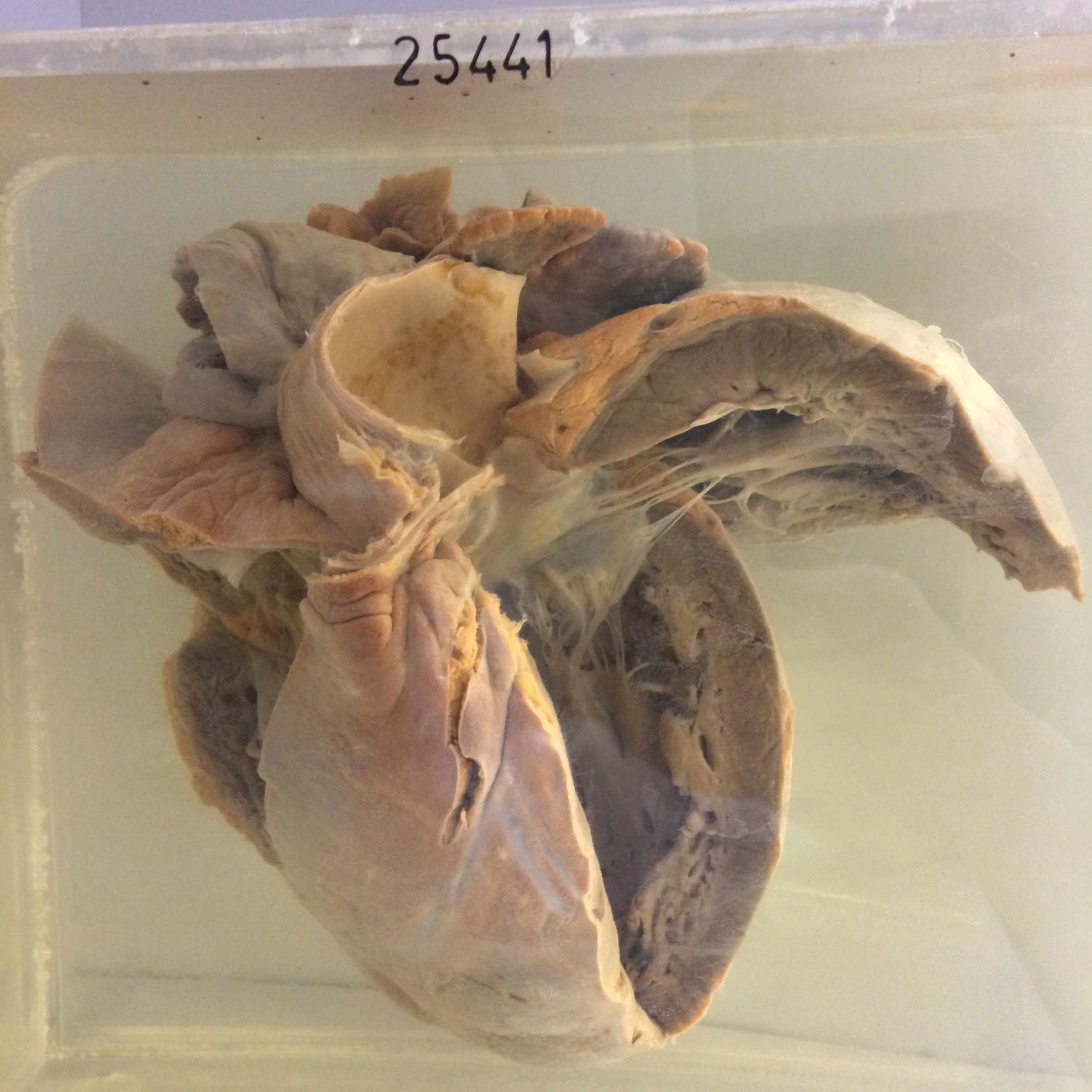
The specimen consists of the heart which is very greatly enlarged. It weighed 715 gm. There is marked dilatation of the left ventricle. The aortic and mitral valves appear grossly normal. The left atrium is slightly hypertrophied but not obviously dilated. The right ventricle is also markedly hypertrophied and moderated dilated. The pulmonary and tricuspid valves appear normal. The right atrium is greatly dilated and hypertrophied. The pericardial surface appears essentially normal. Histology shows irregular patches of hypertrophied muscle fibres. Many muscle nuclei show polar masses of lipofuscin pigment. There are patches of interstitial fibrosis and oedema, but leucocytes are very scanty in the septa.