12481 PROGRESSIVE PRIMARY TUBERCULOSIS
This 18 year old girl was a student nurse who had an episode of pleurisy and was found to have an opacity in the left lower lobe. Bronchoscopy was negative but bronchogram showed a filling defect in a lower lobe bronchus. The lobe was resected.
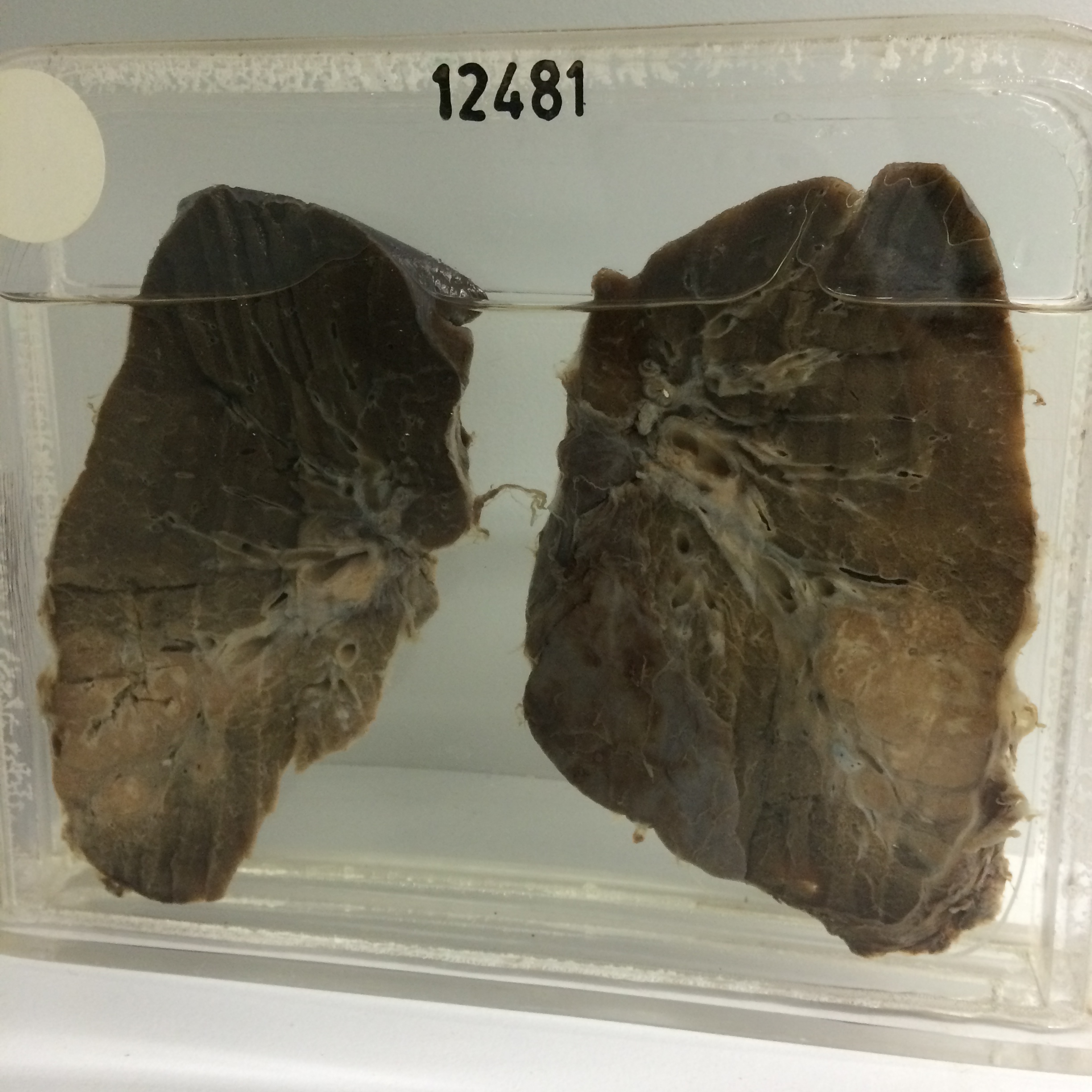
The specimen shows a large area of primary caseous pneumonia measuring 4 x 3 cms extending out to the pleura near the base of the lobe. Fibrous encapsulation is quite well developed and there is no excavation. The overlying pleura is thickened and remnants of fibrous adhesions can be seen. Hilar nodes were found at operation to be caseous. Histology shows caseous necrosis with typical epithelioid cells and Langhan’s giant cells. Acid-fast organisms are present.