14338 EXCAVATED PRIMARY CARCINOMA
This 66 year old man was admitted to the R.A.H. 4 months ago with difficulty in breathing and cough with copious sputum but no haemoptysis. X-ray showed carcinoma of the lung and radiotherapy was begun. A month later there was haemoptysis of 300 mls of blood and he complained of general severe weakness. On examination he was wasted, the left side of the chest was flattened with limited movement and was dull to percussion in the midzone. Haemoglobin 10 g%. X-ray showed a large cavity in the left midzone. He continued to have haemoptysis of about 200 mls/day until the day of his death when he had a further haemoptysis of 600 ml of blood and died within 10 minutes. At postmortem there was a massive primary tumour in the left lung and a solitary metastasis in the left suprarenal.
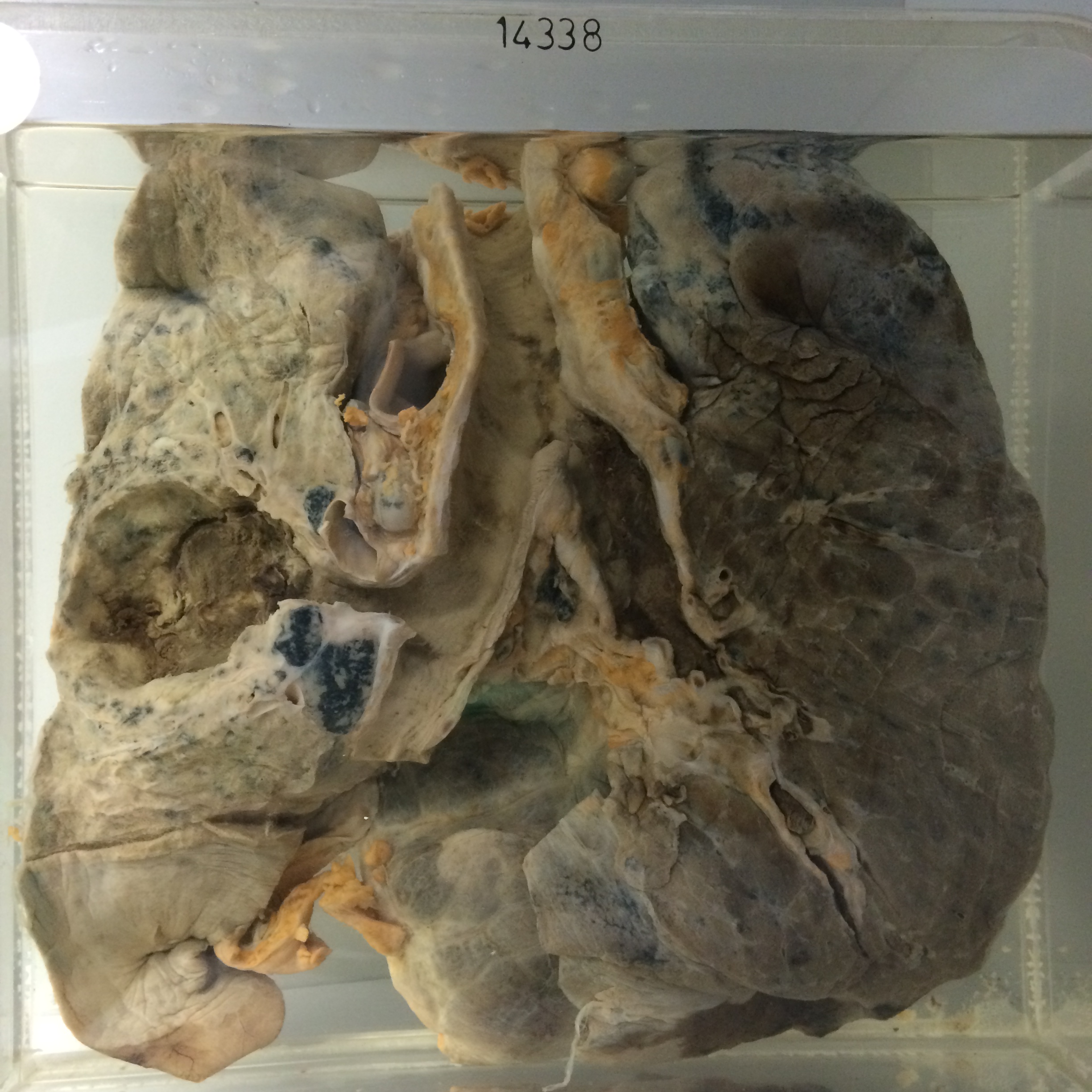
The specimen consists of the lungs and respiratory passages cut to show a large excavated primary carcinoma 6 cms in diameter in the left midzone. Necrotic tumour tissue and recent blood clot are present on the wall of the cavity which communicates by a broad opening into a major bronchus. The surrounding lung shows ill defined grey pneumonic consolidation. There is a mass of recent blood clot in the right main bronchus extending into the intrapulmonary bronchi, and the right lower lobe shows patches of acute haemorrhagic consolidation. Lymph nodes above the major bronchi and on each side of the trachea are heavily involved by tumour. Histology showed anaplastic carcinoma with pleomorphic cells of medium size, tending to form solid acini and occasional tubules.