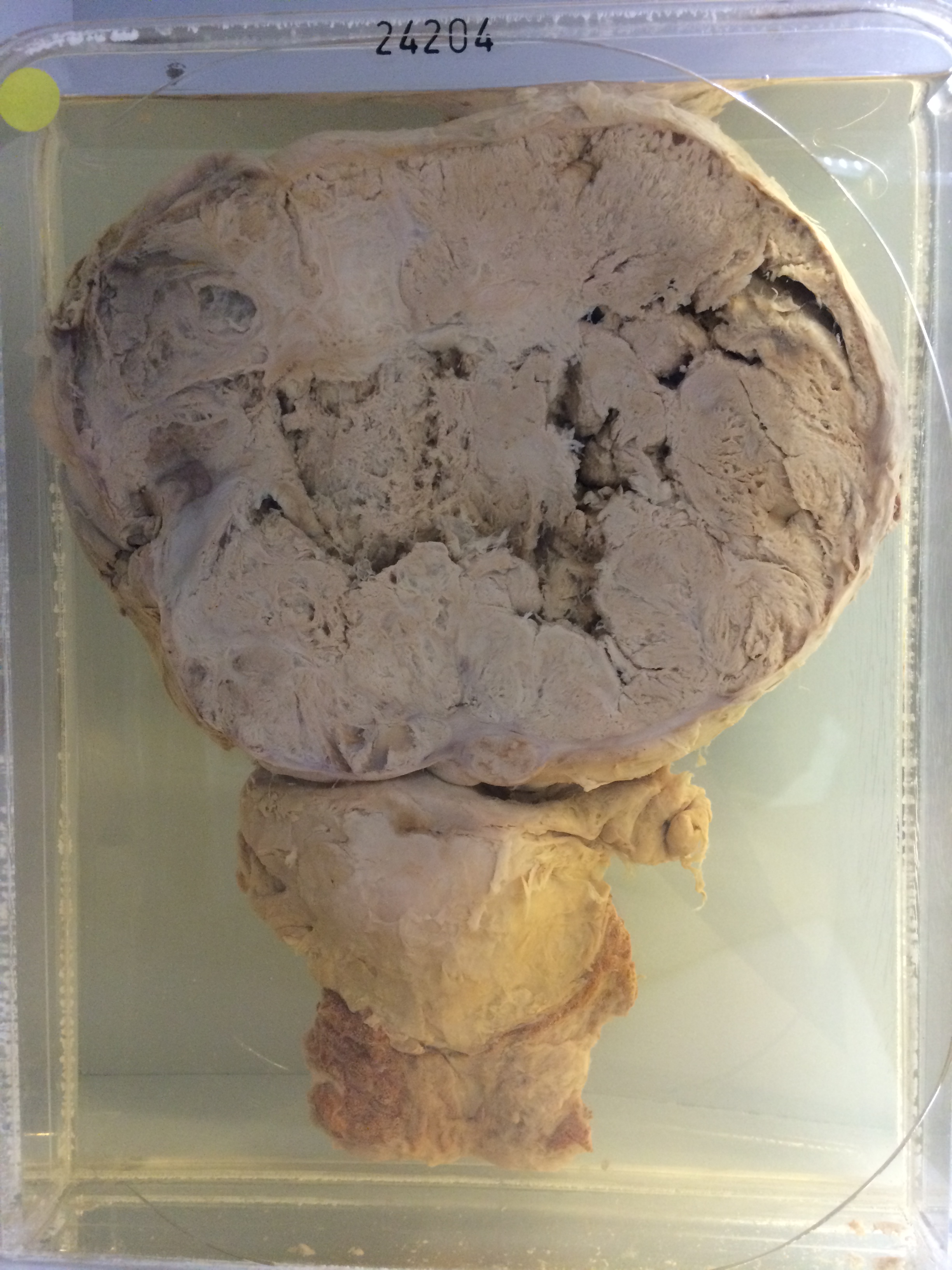
24204 OVARIAN CARCINOMA AND CARCINOMA OF THE UTERINE BODY
The patient was a 59-year old woman who was admitted 2 days before her death with a 10-day history of lower abdominal heaviness. There had been slight blood loss PV. Examination revealed a tense abdomen with ascites and a large mass in the pelvis rising to the umbilicus.
The specimen shows uterus, fallopian tubes and ovaries. Coronal section through the uterus shows carcinomatous tissue filling its cavity and protruding from the cervix. Arising from the ovary is a large gelatinous tumour 20 cms in diameter with much haemorrhage and a central necrotic cavity (on the back of the specimen). Histology shows that the tumour in the ovary is an intracystic papillary columnar celled adenocarcinoma secreting a little mucin in places. The uterine tumour is also an adenocarcinoma with patches of squamous metaplasia. An abdominal lymph node contains secondary carcinoma consisting of large eosinophilic cells forming abortive tubules with some epidermoid features in places.