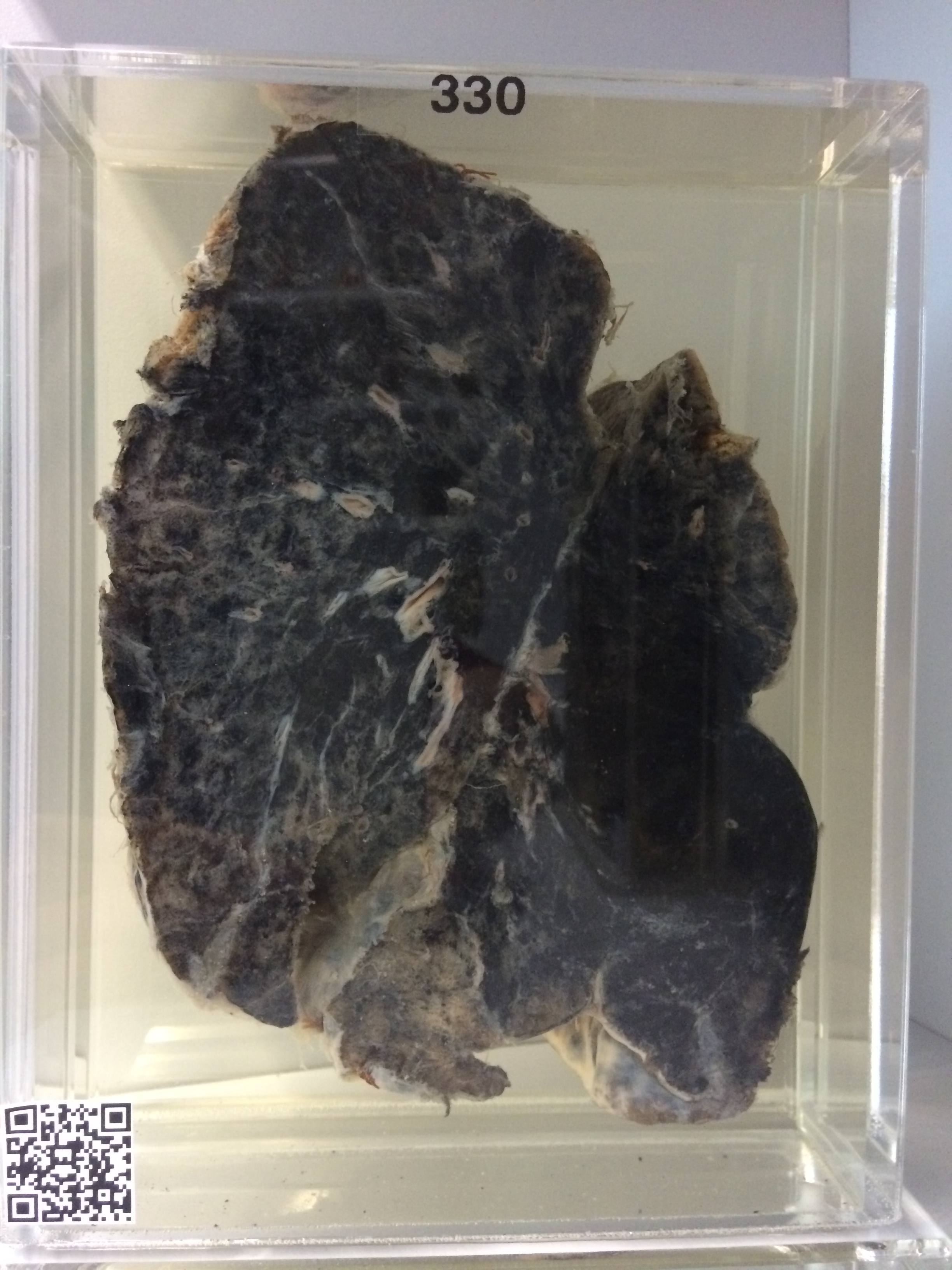
330 SILICOSIS
No clinical information is available.
The specimen is of the left lung which is contracted and fibrosed. The cut surface shows massive anthraco-silicosis which is involving particularly the posterior portion of the lower lobe, where there is a large mass of conglomerate silicosis about 5 cms in diameter. In the upper lobe the process is nodular, with discrete black pigmented masses up to 1 cm in diameter. The intervening lung is emphysematous. The pleura shows dense fibrous adhesions and there is a little acute fibrinous inflammatory exudate at the base.
Last modified: Thursday, 3 August 2017, 9:35 AM