20522 PRIMARY HEPATOMA
The patient was a woman aged 51. Four years previously the gall bladder had been removed. Further investigations suggested either hepatoma or hemangio-endothelioma. The liver biopsy was negative. She was treated conservatively as a cryptogenic cirrhosis and died with gross ascites, jaundice and cardiac failure. At postmortem large venous anastomoses were present beneath the skin over the right lower thoracic region leading downwards towards the umbilicus. There was massive ascites. The liver was of normal size but irregularly nodular. There were gross adhesions to the diaphragm and large venous sinuses were visible ramifying in the adhesions. The hepatic veins and vena cava were normal. Lymph nodes in the porta hepatis were obviously involved by tumour.
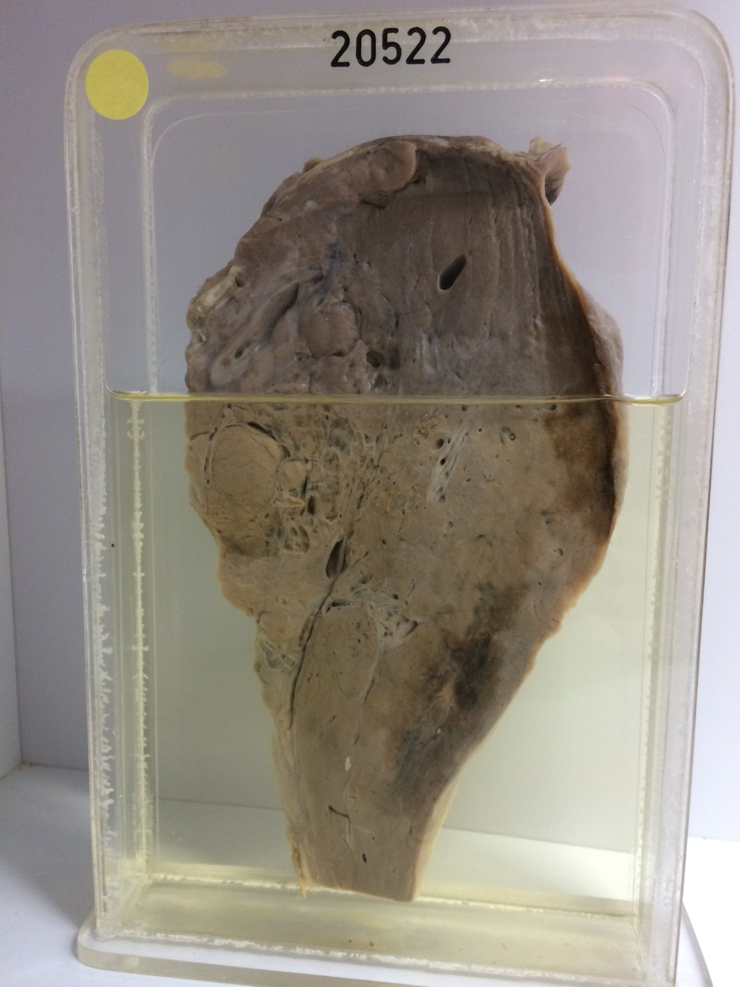
The specimen is a slice of the liver showing massive replacement by lobular confluent masses of pale tumour tissue interspersed with dense fibrous septa. Large spaces and venous channels ramify throughout the tumour. There are two large areas of haemorrhagic congestion beneath the capsule. There is marked hyaline fibrous thickening and nodularity of the overlying capsule. Histology shows a tubular columnar-celled adenocarcinoma with very large sinusoidal veins as a prominent feature.