24755 MASSIVE INTRAMURAL HAEMATOMA OF THE OESOPHAGUS
The patient was a man aged 69. Carcinoma of the tongue first developed 9 years previously and thereafter he was admitted on many occasions for excisions of leukoplakia and recurrent carcinoma. Three months before his death hard fixed nodes were present on the right side of the neck and were irradiated. Thereafter he steadily deteriorated and dysphagia and dementia developed shortly before his death. As postmortem there was massive secondary deposition on the left side of the neck.
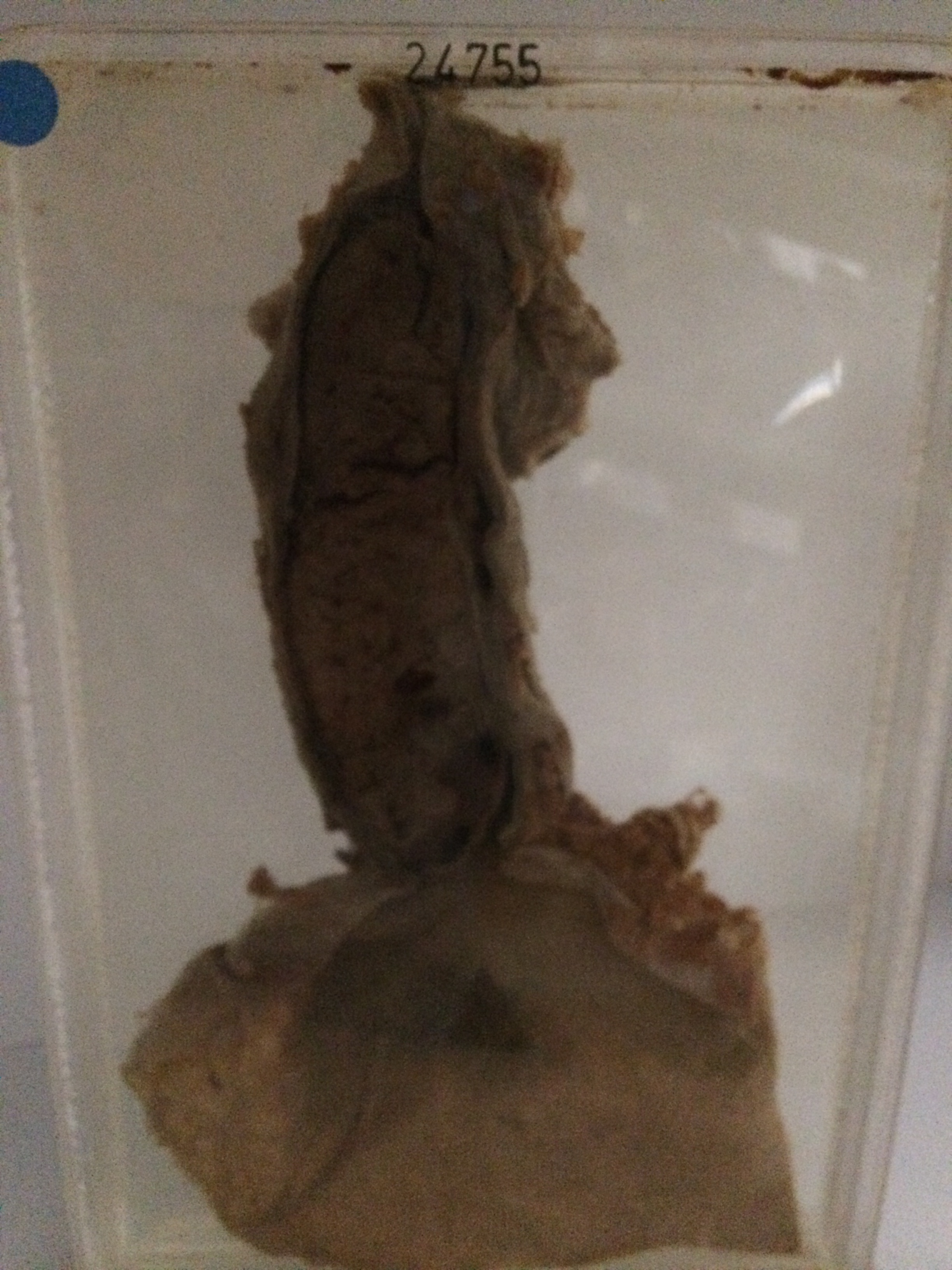
The specimen shows the lower 15 cms of the oesophagus and the upper half of the stomach. There is a most unusual long intra-mural haematoma measuring 14 x 4 x 5 cms in the lower oesophagus, extending to the cardiac entrance. The original lumen of the oesophagus can be seen on the right as a narrow somewhat tortuous channel. Histology shows an acute inflammatory reaction involving all layers and a massive antemortem thrombus closely adherent to the underlying muscular mucosa.